
Diaphragm openings, contents and borders
Read the draft explanation
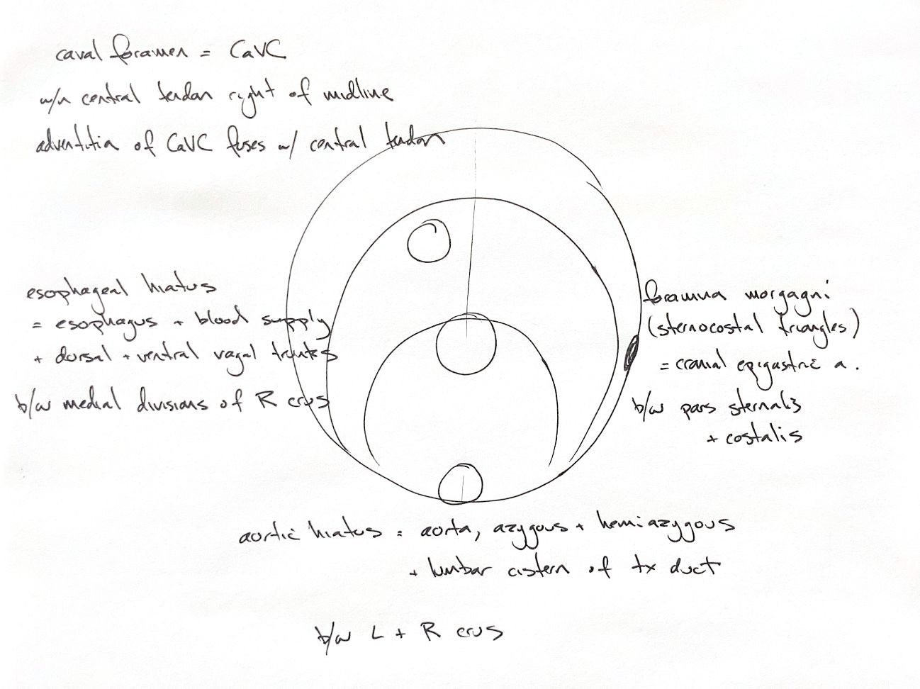
Use the three main openings as a ventral-to-dorsal map. The caval foramen lies in the central tendon, right of the median plane. It carries the caudal vena cava. The vein’s adventitia fuses with the tendon, so this is not a loose gap around the vessel.
The esophageal hiatus lies farther dorsally, between the two medial divisions of the right crus. It carries the esophagus, its blood supply, and the dorsal and ventral vagal trunks. Both named divisions belong to the right crus.
The aortic hiatus is most dorsal. The original places it between the left and right crura. More precisely, lumbar vertebral bodies form its dorsal border, while extensions of the two crural tendons bound it ventrally. The aorta and thoracic duct pass here. The original and book also list azygos and hemiazygos veins; their presence and arrangement must follow the individual anatomy, rather than a compulsory paired-vein template.
Keep the original “lumbar cistern of the thoracic duct” cue. The cisterna chyli is the abdominal lymphatic reservoir that narrows into the thoracic duct at this region. Its shape and extent vary. This continuity explains the different labels without turning the entire cistern into a short tube inside the hiatus.
The smaller paired foramina of Morgagni are the sternocostal triangles. They lie between pars sternalis and pars costalis and transmit the cranial epigastric arteries, which continue from the internal thoracic arteries.
References
Chris Wood’s drawing; Johnston & Tobias, Veterinary Surgery: Small Animal, 2nd ed., chs 85–86, Fig 85.1; University of Minnesota dog/cat anatomy guide; Baum, The Lymphatic System of the Dog; Birch et al., Vet Radiol Ultrasound 2014, PMID23941469 (complete abstract).