
Abdominal-wall composition from diaphragm to pelvic canal
Read the draft explanation
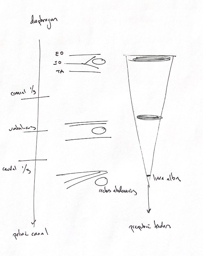
Follow the rectus abdominis from the diaphragm toward the pelvic canal. Its sheath changes along the way. An aponeurosis is a broad, flat tendon. Here, the aponeuroses of external abdominal oblique, internal abdominal oblique and transversus abdominis pass around rectus abdominis before joining at the linea alba.
External oblique stays superficial to rectus throughout. In the cranial third, internal oblique splits, with fibres passing superficial and deep to rectus. Transversus stays deep. Around the umbilical region, internal oblique has moved superficial, while transversus still passes deep. In the caudal third, all three aponeuroses pass superficial to rectus.
These are schematic regional comparisons, not exact boundaries measured in one patient. They show the muscle aponeuroses. Transversalis fascia and peritoneum still line the inner abdominal wall, even where no deep aponeurotic sheath is drawn.
The linea alba is the midline join between the left and right rectus muscles. It is broadest cranially. Caudal to the umbilicus, it narrows and thickens, then blends with the prepubic tendon at the pubis. Keep the original tapering strip beside the three wall sections: moving caudally changes both the sheath arrangement and the width of the midline join.
References
Chris Wood’s drawing; Johnston & Tobias, Veterinary Surgery: Small Animal, 2nd ed., chs 84 and 86, Figs 84.2 and 86.2; University of Minnesota dog/cat abdominal-wall anatomy guide.