
Vaginal embryology: cloacal partition and paramesonephric fusion
Read the draft explanation
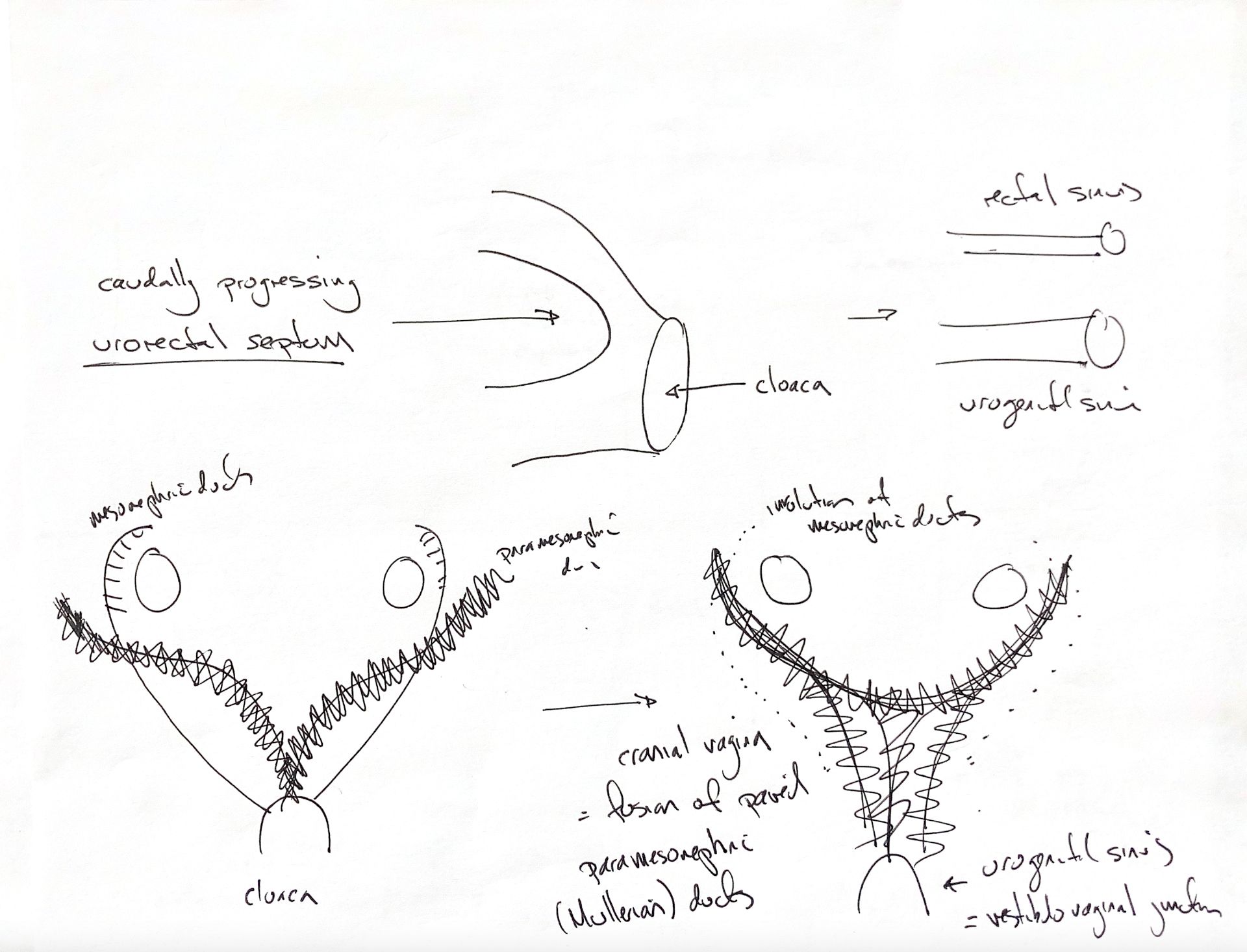
The cranial vagina forms when the paired paramesonephric, or Müllerian, ducts fuse. Keep that answer beside the two changes in the drawing.
First, the early digestive and urogenital pathways share a chamber called the cloaca. The urorectal septum progresses caudally and separates them. The dorsal part becomes the rectum and anal canal. The ventral part is the urogenital sinus. The original “rectal sinus” label is shown here as the rectum and anal canal.
Next, follow the two duct systems. During female development, the mesonephric, or Wolffian, ducts regress. The dotted lines retain their former paths. The paramesonephric ducts persist, and their caudal portions fuse into one midline tube. This fused part forms the cranial vagina.
The fused tube meets the more caudal urogenital sinus at the vestibulovaginal junction, also called the cingulum. The caudal urogenital sinus forms the vestibule. The junction is where the two regions meet; it is not another name for the whole sinus.
References
Chris Wood’s drawing; Johnston & Tobias, Veterinary Surgery: Small Animal, 2nd ed., ch 110 and Fig 110.1; University of Minnesota veterinary developmental-anatomy resources. Cornell’s canine embryonic atlas also records female paramesonephric ducts beside regressed mesonephric ducts.