
Meniscal hoop stress and release
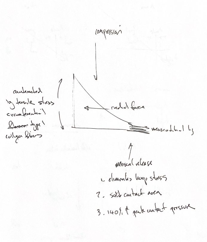
The study reports about a 41% increase in peak pressure, or 141% of baseline. The handwritten 140% increase overstates the change.
Read the draft explanation
Compression pushes the wedge-shaped meniscus outward. Its strong, mainly type I collagen bundles run around the rim. They develop tensile hoop stress, which restrains radial extrusion. The cranial and caudal meniscotibial ligaments carry these forces to the tibia. The meniscal body, peripheral rim and attachments work as one functional unit.
Cutting the caudal meniscotibial ligament disrupts this load path. The original meniscal-release cue describes loss of effective hoop-tension load sharing. It does not mean every collagen fibre has zero stress.
The numerical cue needs a source correction. In eight canine cadaveric limbs with CCL-deficient stifles stabilised by TPLO, the published abstract reports mean medial contact area falling from 145 ± 24 to 71 ± 25 mm²—about 50%. Mean peak pressure rose from 2.9 ± 0.4 to 4.1 ± 0.5 MPa. Those means imply about a 41% increase, or 141% of baseline. The original note and second-edition book say a 140% increase. That conflicts with the cited abstract and should not be repeated as the same quantity.
The limbs were loaded at 30% body weight with the stifle at 135°. These are ex vivo contact mechanics, not a fixed effect in every living dog or a clinical complication rate. Preserving functional meniscal tissue supports load sharing.
References
Chris Wood’s ACVS notes; Johnston & Tobias, Veterinary Surgery: Small Animal, 2nd ed., ch. 38 and 61, supplied PDF pp. 1535, 2516–2517 and 2521–2524; Pozzi, Kim & Lewis, Vet Surg 2010;39:489–495, primary abstract, doi:10.1111/j.1532-950X.2010.00662.x. Full primary article not obtained.