
CCL bands, meniscal tear forms and Sharpey-fibre attachment
Read the draft explanation
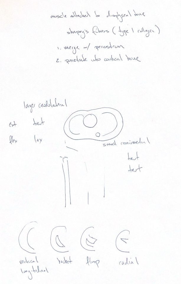
Sharpey's fibres help anchor a fibrous attachment to bone. These dense collagen bundles are predominantly type I. At muscle attachments to the diaphysis, the tendon fibres merge with the periosteum and penetrate deeply into cortical bone. An enthesis is an attachment site. Fibrocartilaginous entheses have a different transition, with no periosteum at that interface.
For the canine CCL, learn both band names and both joint positions. In the classical gross description, the caudolateral band is larger and the craniomedial band is smaller. The CM band stays taut in flexion and extension. The CL band is taut in extension but lax in flexion. Both help check cranial tibial translation, with the CM band providing the primary check across these positions.
That is a useful gross model, but fibre behaviour is more complex. A later Beagle cadaver study measured larger CM attachment footprints than CL footprints. The size cue in the gross description is not a universal rule for measured attachment area.
The four tear sketches keep the meniscus in the same orientation. A vertical longitudinal tear follows the circumferential fibres and extends between the tibial and femoral surfaces. It can be complete or incomplete. A bucket-handle tear is a displaced longitudinal tear whose fragment remains attached at both ends. A flap forms when one end tears free. A radial tear runs from the inner free edge toward the peripheral rim.
These four forms are the selected recall set, not the whole meniscal classification. The attachment sketch is a separate connective-tissue concept; it does not make every cruciate attachment a fibrous periosteal enthesis.
References
Johnston and Tobias, Veterinary Surgery: Small Animal, 2nd ed., Chapters 38 and 61; Tanegashima et al., 2019. University of São Paulo histology guide.