
Tarsal supply and central tarsal fractures
Read the draft explanation
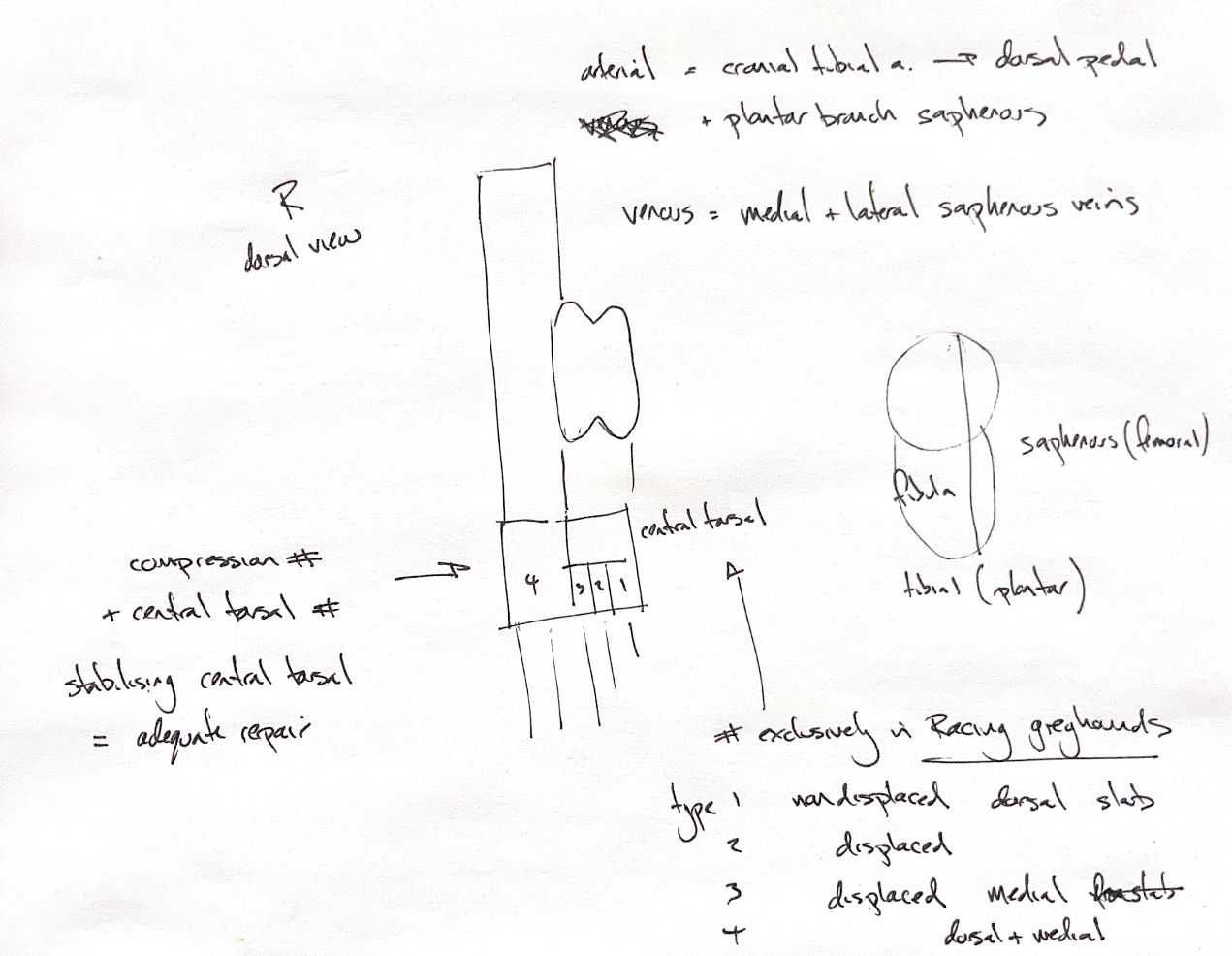
The tarsus has two main arterial routes. The cranial tibial artery becomes the dorsal pedal artery at the tarsocrural joint, then gives rise to dorsal metatarsal arteries. It supplies the dorsal and lateral tarsus. The plantar branch of the saphenous artery passes through the tarsal canal and supplies the medial and plantar regions. Venous blood drains through the medial and lateral saphenous veins.
The sciatic nerve gives rise to the tibial and common fibular, or peroneal, nerves. The tibial nerve divides into medial and lateral plantar nerves. The common fibular nerve divides into superficial and deep branches. The saphenous nerve comes from the femoral nerve and provides dorsomedial cutaneous supply.
The central tarsal bone acts as a buttress. Its fracture can allow collapse and plantar flexion, with added strain on T4 or the calcaneus. T4 compression fractures often accompany central-tarsal injury. Restoring central-tarsal alignment and stability is usually enough to allow the accompanying T4 fracture to heal. Other associated injuries still need assessment.
Central-tarsal fractures occur almost exclusively in racing Greyhounds and usually affect the right hock. Other breeds and either side can be affected. Using the supplied second-edition scheme, type I is a nondisplaced dorsal slab; type II, a displaced dorsal slab; type III, a large displaced medial fragment; type IV, both dorsal and medial slabs; and type V, a comminuted fracture. Older schemes describe type III as dorsal and/or medial slabs.
References
Johnston & Tobias, Veterinary Surgery: Small Animal, 2nd ed., ch. 64; Boudrieau et al., JAVMA 1984;184:1486–1491; Evans & de Lahunta, Miller’s Anatomy of the Dog, 4th ed. (anatomy cited in the supplied text).