
Tarsal collateral ligaments
Read the draft explanation
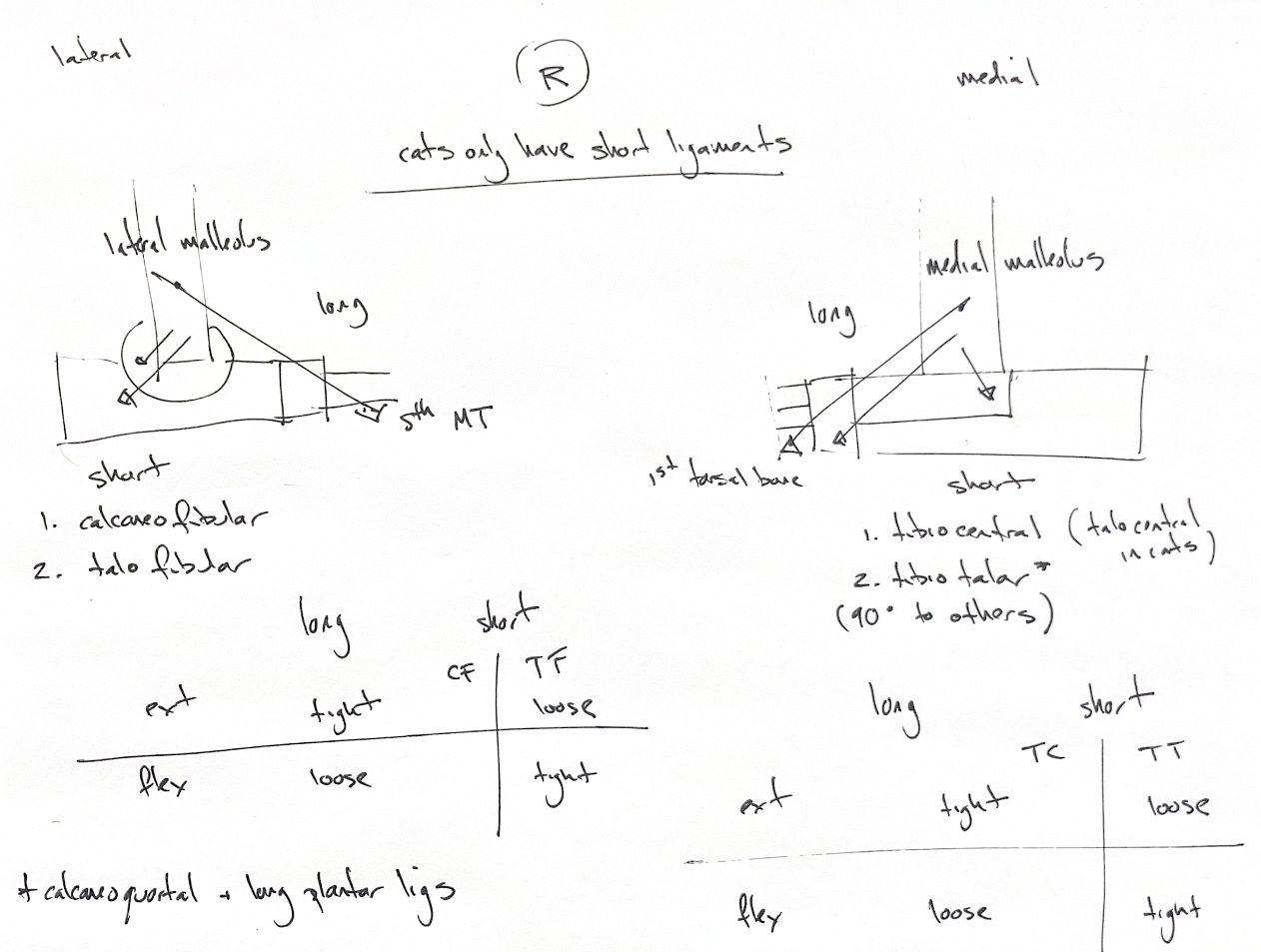
The dog’s collateral ligaments have long and short components. Laterally, the long part runs from the lateral malleolus toward metatarsal V. Its short parts are calcaneofibular and talofibular. Medially, the long part runs from the medial malleolus to T1, with a firm central-tarsal attachment. The short parts are tibiocentral and tibiotalar. The tibiotalar course lies about 90° to the other medial parts.
In extension, both long parts and the short medial tibiocentral part are tight. They loosen in flexion. The short tibiotalar and talofibular parts tighten in flexion and are loose in extension. The calcaneofibular part supports extension, but it also contributes to static stability in flexion. Do not equate a tension pattern with the whole contribution to stability. In a canine cadaver study, medial tibiotalar and lateral calcaneofibular parts were important in both positions. The capsule, malleoli and ligament complex work together.
Cats have only short collateral parts. Medially, later cadaver descriptions identify tibiotalar and tibiocentral parts. The tibiocentral band runs from the medial tibial malleolus to the dorsomedial central-tarsal process. The supplied second-edition text calls this straight feline band “talocentral”; this name differs from the later description. Laterally, cats have calcaneofibular and talofibular parts.
The calcaneoquartal and long plantar ligaments add plantar support. Keep these distinct from the collateral parts.
References
Johnston & Tobias, Veterinary Surgery: Small Animal, 2nd ed., ch. 64; Aron & Purinton, Vet Surg 1985;14:173–177; Luescher et al., Vet Surg 2020;49:390–400, doi:10.1111/vsu.13366.