
Right carpus: recall map and labelled reference views
Read the draft explanation
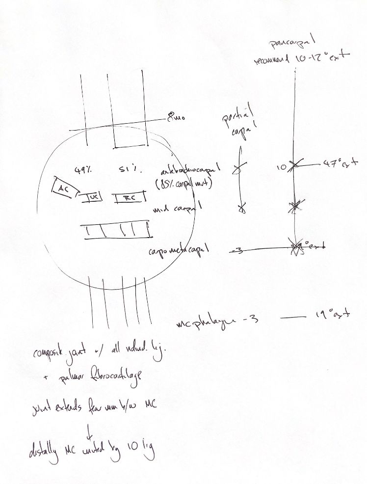
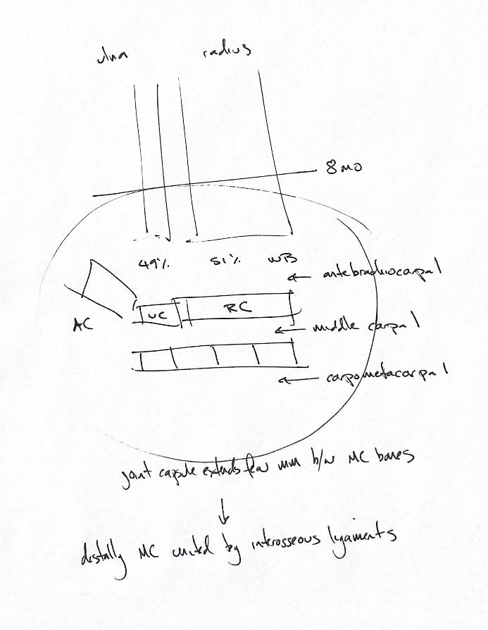
Start with the right carpus as two rows of bones. RC, or CR in the reference figures, is the large intermedioradial carpal. UC is the ulnar carpal. AC projects palmarly. The four numbered carpals form the distal row. The three main levels are antebrachiocarpal, middle carpal and carpometacarpal. Carpometacarpal is distinct from the metacarpophalangeal joint farther down each digit.
The capsule sends recesses between the proximal metacarpals. The original “few millimetres” cue is approximate; recess extent varies. Interosseous metacarpal ligaments unite the bones farther distally. The antebrachiocarpal cavity is separate from the communicating middle carpal and carpometacarpal cavities. Palmar fibrocartilage and short ligaments add support.
The original 49% ulna/51% radius cue needs a change of location. Mason’s force study measured the elbow surfaces in 12 cadaveric right forelimbs, not the carpus. Radius carried 51–52% under 50–200 N loads. The 8-month closure cue is also approximate. The book gives 222–250 days in Beagles; timing varies with breed and physis.
The book summarises 10–47° of antebrachiocarpal extension and −3° to 19° at the metacarpophalangeal joint. Gait and measurement methods affect these angles. Figure 55.6’s plotted signs conflict with the book’s text convention. The traditional pancarpal fusion target is 10–12° of extension. Partial carpal fusion preserves the antebrachiocarpal level when that joint is intact.
The reference sequence keeps deep palmar, superficial palmar, dorsal and lateral views separate. The palmar and dorsal figures show the left paw in the supplied text. Use the sesamoid pairs to find the intersesamoidean and cruciate ligaments. Follow digits II–V from MCP to PIP and DIP collateral ligaments, then the dorsal elastic ligaments near the claw. Digit I has two phalanges; the other four have three. The flexor retinaculum and palmar fibrocartilage are different structures.
References
Chris Wood’s notes; Johnston & Tobias, Veterinary Surgery: Small Animal, 2nd ed., ch.54–55, figs55.1/3–5; Mason et al. 2005 AJVR66:132–135; Klußmann et al. 2024, doi10.1111/ahe.13026.