
Primary and secondary hip stabilisers
Read the draft explanation
The hip stays stable through primary supports and a second layer of soft-tissue and muscle support.
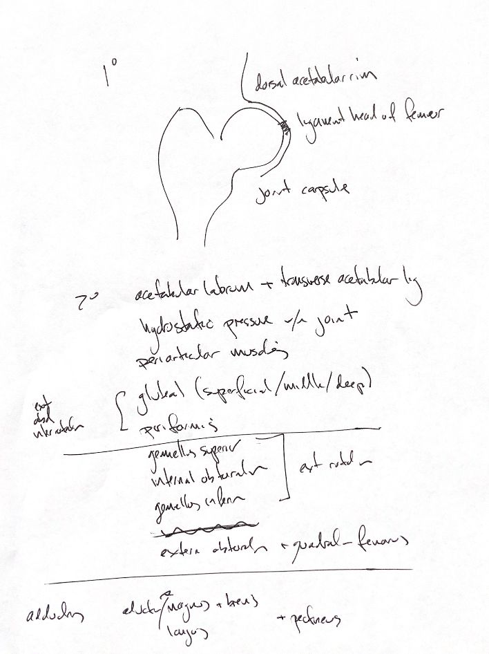
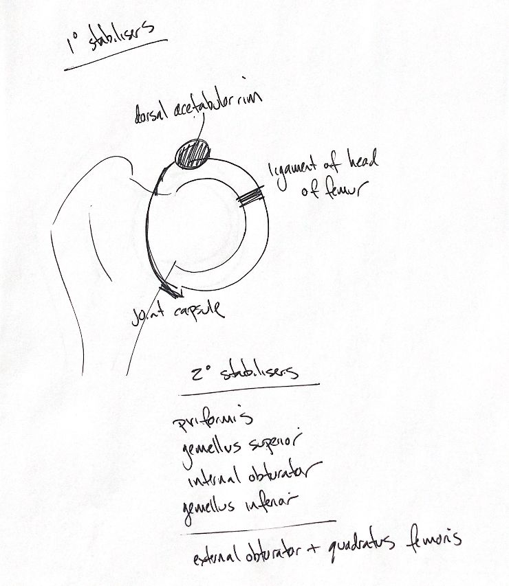
The primary stabilisers are the dorsal acetabular rim, the ligament of the head of the femur and the joint capsule. The bony rim supports the head dorsally. The head ligament runs from the fovea capitis to the acetabular fossa. The capsule links the acetabular rim region to the femoral neck.
The acetabular labrum adds secondary support. This thin fibrocartilaginous band extends laterally from the rim. Across the ventral acetabular notch, it forms the transverse acetabular ligament. Hydrostatic pressure within the fluid-filled joint also helps stabilise it.
Periarticular muscles cross the hip and add support. It helps to learn these muscles in groups. The superficial, middle and deep gluteals, with piriformis, form the extension and abduction group. The gluteal group also contributes to medial rotation; this group cue does not mean every muscle has identical actions.
The lateral rotators are the gemelli, internal obturator, external obturator and quadratus femoris. The note calls the gemelli superior and inferior. In canine anatomy, they are usually named together as the gemelli, with paired bellies around the internal obturator tendon.
For adduction, retain adductor magnus et brevis, adductor longus and pectineus. Adduction moves the limb toward the midline; abduction moves it away.
References
Johnston and Tobias, Veterinary Surgery: Small Animal, 2nd ed., Chapter 57; University of Minnesota canine dissection anatomy.