
Osteoarthritis catabolic signalling
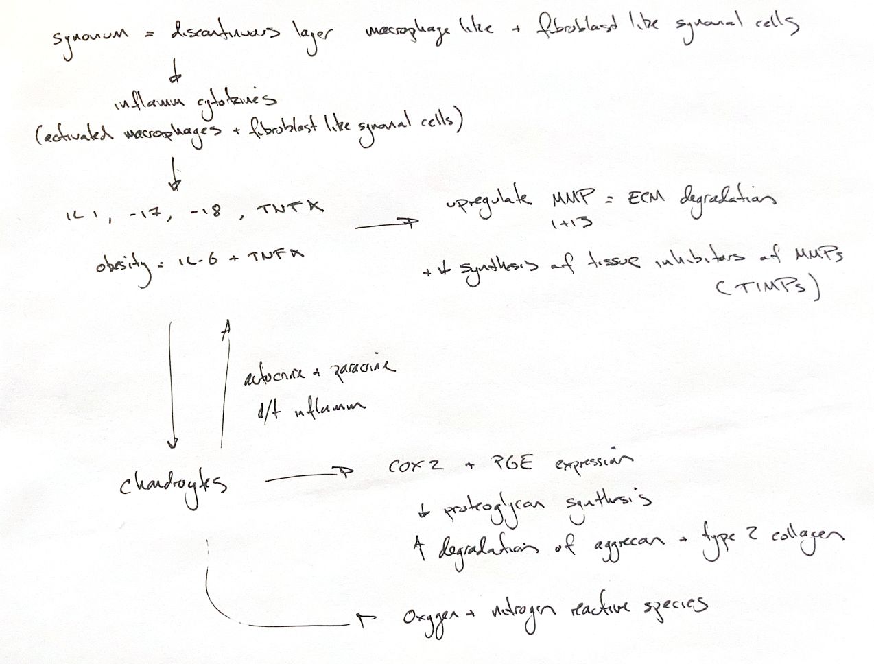
Read the draft explanation
The synovium and cartilage communicate during osteoarthritis. Activated macrophages and fibroblast-like synovial cells release inflammatory mediators. IL-1, IL-17, IL-18 and TNF-α can increase matrix metalloproteinases (MMPs) and other proteases while tissue inhibitors of metalloproteinases, or TIMPs, decrease in the simplified catabolic model. Expression varies with site and stage. The balance shifts toward matrix breakdown. MMP-1 and MMP-13 can damage the type II collagen network.
Chondrocytes also make cytokines. Autocrine signals act on the cell that released them. Paracrine signals act on nearby cells. This feedback can sustain inflammation. In canine obesity, IL-6 and TNF-α may increase, but cohorts differ. One study found no difference from normal-weight dogs. A systemic contribution to OA remains proposed.
The chondrocyte response includes cyclooxygenase-2 (COX-2) and prostaglandin E₂ (PGE₂) production. In human OA cartilage explants, PGE₂ reduced proteoglycan synthesis and promoted aggrecan and type II collagen breakdown. These effects depend on the model and receptor context. They are not a universal canine response.
Reactive oxygen and nitrogen species add another pathway. Oxidative stress can promote chondrocyte apoptosis. Cytokines also switch on inducible nitric oxide synthase (iNOS). This enzyme produces nitric oxide. Excess NO can inhibit matrix synthesis, activate MMPs and promote apoptosis. Yet NO can also protect cartilage: blocking its production can increase protease activity and proteoglycan loss. Its role depends on context.
References
Chris Wood's ACVS notes; Johnston & Tobias, Veterinary Surgery: Small Animal, 2nd ed., ch.68, Fig.68.3, supplied PDF2924,2926–2929. Attur et al., J Immunol 2008;181:5082–5088, doi:10.4049/jimmunol.181.7.5082. Piantedosi et al., Vet J 2016;216:72–78.