
Osteoarthritis tissue changes
Read the draft explanation
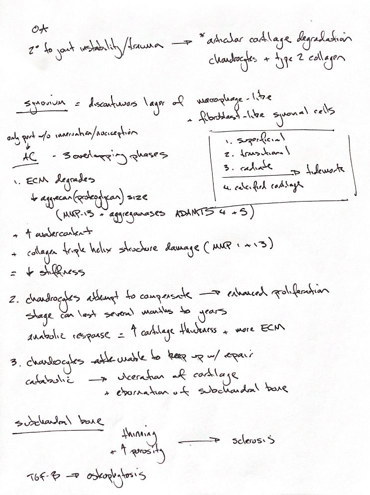
Canine osteoarthritis usually follows a joint problem such as instability or trauma. Articular cartilage contains chondrocytes in a type II collagen matrix. Its superficial, transitional and radiate zones sit above the tidemark and calcified cartilage. Normal cartilage has no nociceptors. The synovium is a discontinuous lining of macrophage-like and fibroblast-like cells. Synovitis and capsular fibrosis vary.
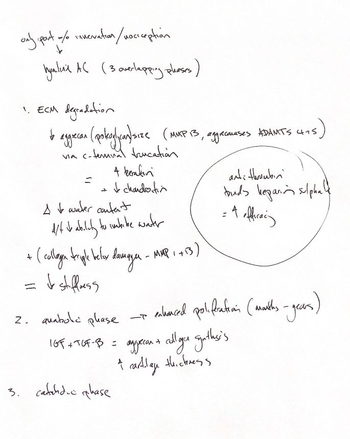
Three overlapping phases help explain cartilage loss. First, matrix metalloproteinases (MMPs) and aggrecanases break down matrix. MMP-13 can cleave aggrecan; ADAMTS-4 and -5 are important aggrecanases. Their relative importance in dogs remains unresolved. MMP-1 and -13 damage the type II collagen triple helix. Early OA cartilage gains water and loses stiffness.
The original water-decrease cue conflicts with this early OA swelling. It fits the separate ageing-aggrecan explanation: C-terminal truncation leaves less chondroitin sulfate and more keratan sulfate, reducing the ability to imbibe water.
Next, chondrocytes proliferate and make more matrix. Insulin-like growth factor (IGF) and transforming growth factor-β (TGF-β) can stimulate aggrecan and collagen synthesis. Cartilage may thicken, and this repair attempt can last months to years. Eventually, repair cannot keep up. Cartilage ulcerates, exposing polished subchondral bone: eburnation.
In canine cruciate-transection models, subchondral plate thinning and increased porosity precede sclerosis. Nearby cartilage and bone damage do not establish which caused which. TGF-β can induce osteophytes in experimental animals.
A separate GAG cue in the original note concerns anticoagulation. Specific heparan-sulfate sequences bind antithrombin and speed inhibition of coagulation proteases. This is separate from the OA pathway.
References
Chris Wood's ACVS notes; Johnston & Tobias, Veterinary Surgery: Small Animal, 2nd ed., ch.68, supplied PDF2921–2929; ch.7, PDF328. HajMohammadi et al., J Clin Invest 2003;111:989–999, doi:10.1172/JCI15809.