
Osteochondrosis types and sites
Read the draft explanation
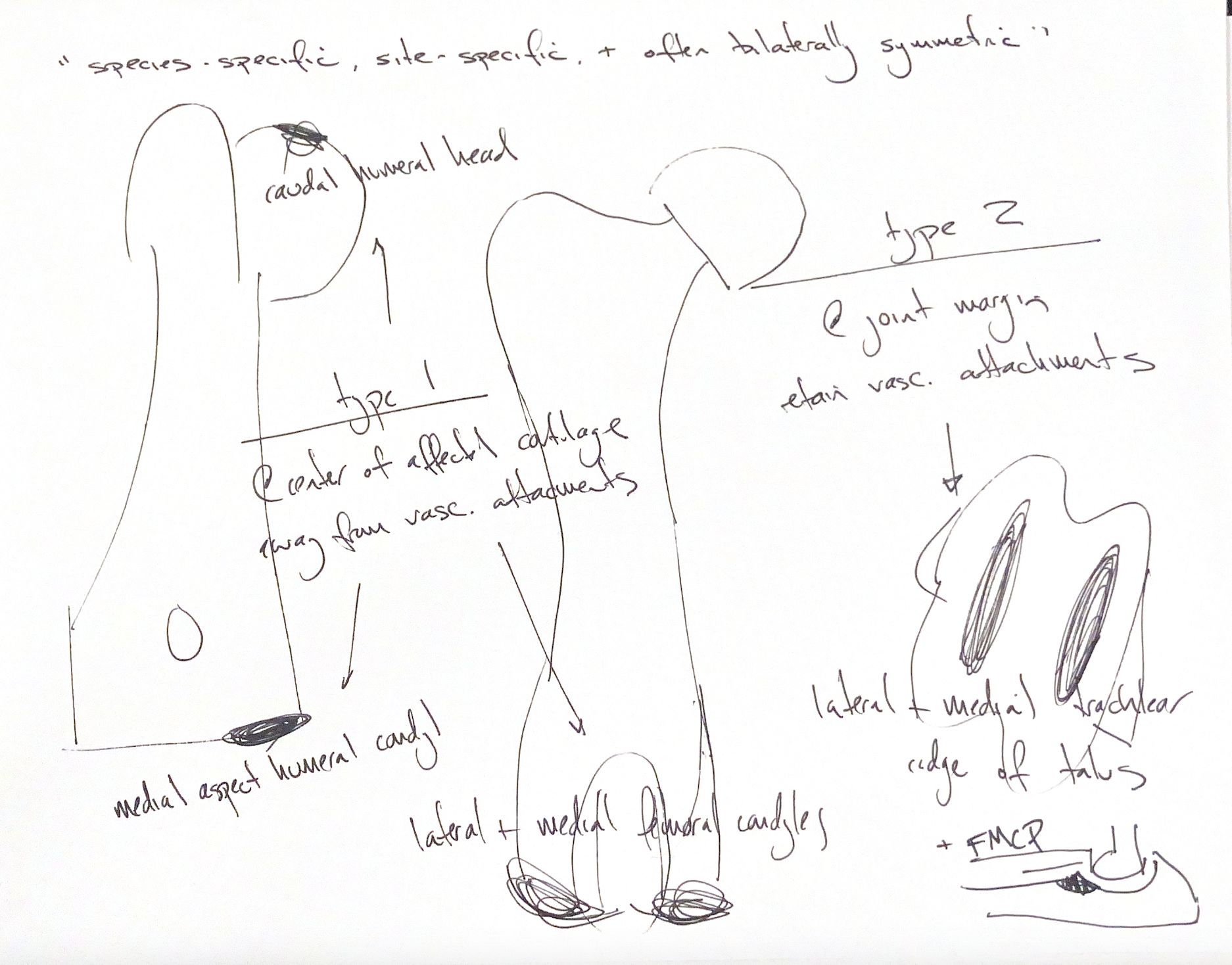
Osteochondrosis disrupts the normal change from growth cartilage to bone. Its lesions favor specific sites in each species and often occur on both sides in a similar pattern.
Olsson and Reiland proposed two anatomical types. The key is where the lesion sits and whether a vascular attachment remains.
Type I lesions sit near the center of an articular surface, away from vascular attachments. In the dog, the main sites in this map are the caudal humeral head, the medial aspect of the humeral condyle, and the femoral condyles. The lateral femoral condyle is affected most often, but the medial condyle can also be involved.
Type II lesions sit at a joint margin and retain a vascular attachment. Examples in this proposed classification are the medial or lateral trochlear ridges of the talus and a fragmented medial coronoid process, or FMCP. Talar lesions attach to the joint capsule and/or collateral ligaments. The medial coronoid process receives vessels through the capsule and/or annular ligament.
FMCP affects the proximal ulna. The tip of the medial coronoid process lies distal to the radial head. Elbow OCD affects the medial humeral condyle. FMCP has a complex pathogenesis; osteochondrosis and mechanical overload are among the proposed mechanisms. Its inclusion in the type II map is a classification proposal. Most knowledge of osteochondrosis pathogenesis comes from work on type I lesions.
References
Johnston & Tobias, Veterinary Surgery: Small Animal, 2nd ed., ch. 73, “Articular–Epiphyseal Cartilage Complex”, and ch. 52, Fig. 52.1; Olsson & Reiland, Acta Radiol Suppl 1978;358:299–306 (classification cited in the supplied textbook).