
Distraction osteogenesis
Read the draft explanation
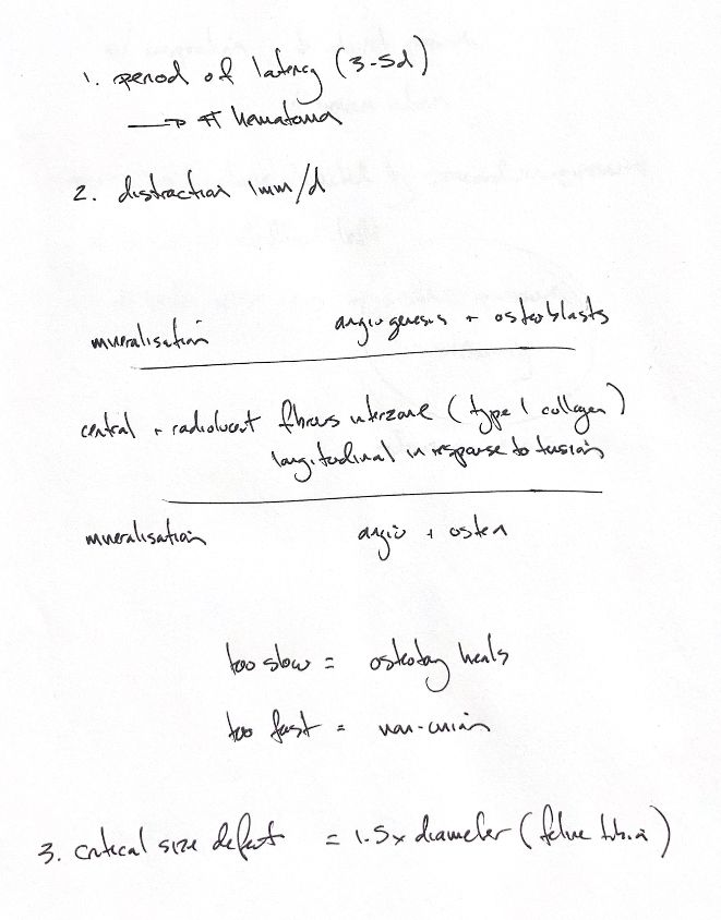
Distraction osteogenesis creates new bone by applying controlled tension. After an osteotomy, stable fixation allows an early haematoma to form. A usual latency is 3–5 days. Age, bone site and healing potential affect that interval.
Distraction then proceeds slowly. About 1 mm/day is a common reference rate, supported by Ilizarov’s canine tibial experiments. The rate is adjusted to the patient and the developing regenerate. Too slow, and the osteotomy may unite before lengthening is complete. Too fast, and the early tissue bridge can be disrupted, leading to nonunion.
Tension aligns the provisional matrix along the direction of lengthening. Fibroblasts and mesenchymal stem cells lay down longitudinal type I collagen in the central fibrous interzone. This zone stays radiolucent during steady distraction. On both sides, blood vessels grow in and osteoblasts place osteoid along the collagen bundles. These flanking zones mineralise and lengthen while the central interzone stays about the same width.
Once distraction stops, the central interzone mineralises during consolidation. The regenerate must mature before it can safely take over the load.
Critical-sized defects depend on the species and bone site. Key’s hypothesis used a segmental defect 1.5× the bone diameter in skeletally mature dogs. The cited feline study tested 5 adult cats with rigidly fixed tibial defects of 1.25–1.52× the diaphyseal diameter. At 12 weeks, there were 4 nonunions and 1 equivocally delayed union. This does not establish a sharp 1.5× cutoff for cats. Species, age, bone site and the local tissues affect how large a defect can heal. Bone transport can create regenerate behind a moving segment to help reconstruct such defects.
References
hand-drawn ACVS Phase One notes; Johnston and Tobias, Veterinary Surgery: Small Animal, 2nd edition, Chapters 39 and 42. Ilizarov, Clin Orthop Relat Res 1989;239:263–285. Toombs et al., Am J Vet Res 1985;46:513–518.