
Stem-cell reservoirs and bone growth factors
Read the draft explanation
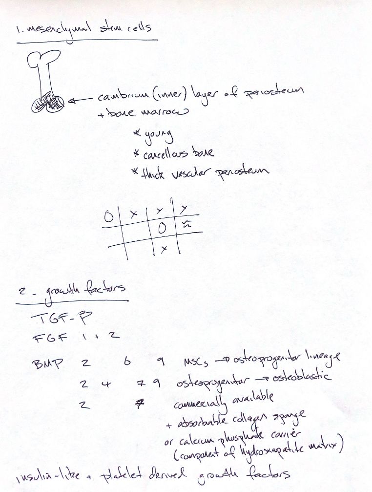
Bone repair needs cells and signals. Mesenchymal stem cells can enter the bone-forming lineage. Your main reservoirs are the cambium, the inner layer of the periosteum, and bone marrow. Young patients, cancellous bone and thick, vascular periosteum provide a favourable cellular environment. Cell numbers fall with age, but stem cells remain throughout life.
The signal list starts with transforming growth factor beta, TGF-β. It acts on several cell types during repair. Fibroblast growth factors 1 and 2, FGF-1 and FGF-2, support cell proliferation and angiogenesis: the formation of new blood vessels. Insulin-like growth factors, IGFs, support skeletal growth and local cell proliferation. Platelet-derived growth factor, PDGF, is released from platelet alpha-granules into the early fracture environment.
Bone morphogenetic proteins, or BMPs, guide osteogenic differentiation. Keep your two-stage memory map: BMP-2, -6 and -9 act on mesenchymal progenitors; BMP-2, -4, -7 and -9 have roles as osteoprogenitors become osteoblasts. The lists are examples, not exclusive pathways. Experimental cell work also found BMP-6 activity in preosteoblastic cells. These models do not establish a guaranteed clinical response in dogs.
Your BMP-2 and BMP-7 availability cue belongs to the textbook's era. The recombinant human proteins are rhBMP-2 and rhBMP-7. The carrier matters because soluble BMP clears rapidly from the local site. Absorbable collagen sponge and selected calcium phosphate materials are carrier examples. Hydroxyapatite is itself a calcium phosphate in bone mineral.
A factor's role in normal repair does not prove that adding it improves healing. The textbook describes inconsistent TGF-β results and limited evidence for exogenous IGF or combined IGF–PDGF benefit.
References
hand-drawn ACVS Phase One notes; Johnston and Tobias, Veterinary Surgery: Small Animal, 2nd edition, Chapters 45 and 48; Cheng et al., JBJS 2003, doi:10.2106/00004623-200308000-00017.