
Nonunion patterns and biological viability
Read the draft explanation
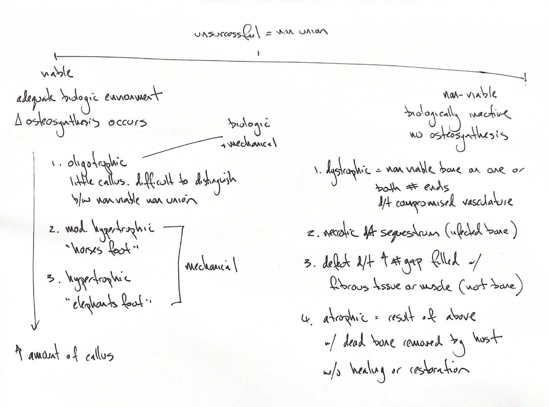
The classic non-union scheme groups fractures by their healing response and bone-end appearance. The names help organise the problem, but morphology alone does not measure blood supply or tissue viability.
In the viable group, some biological response remains. Oligotrophic non-union has little radiographic callus and can resemble a biologically inactive fracture. Mechanical and biological problems may coexist. Moderately hypertrophic non-union has more callus, the “horse’s foot” pattern. Hypertrophic non-union has abundant flared callus, the “elephant’s foot” pattern. Mechanical instability often predominates in these two hypertrophic groups. The callus still fails to bridge the fracture.
The classic nonviable group includes four patterns. Dystrophic non-union has devitalised bone at one or both ends after vascular compromise. The necrotic type contains an infected sequestrum. A sequestrum is dead bone; infection is the feature of the type described here, rather than a requirement for every sequestrum.
Defect non-union has a gap beyond the local capacity to regenerate bone. Fibrous tissue or muscle occupies that gap. Atrophic non-union can follow these failures: bone is resorbed without a matching restorative response, leaving tapered or rounded ends and little callus.
An atrophic image does not prove avascularity. In a study of eight non-unions in six dogs, radiographic predictions of viability underestimated the vascularity found on histology. Keep the classic pattern names while assessing the actual biological and mechanical problem.
References
hand-drawn ACVS Phase One notes; Johnston and Tobias, Veterinary Surgery: Small Animal, 2nd edition, Chapter 45; Risselada et al., AJVR 2006, doi:10.2460/ajvr.67.8.1354.