
The five phases of secondary healing
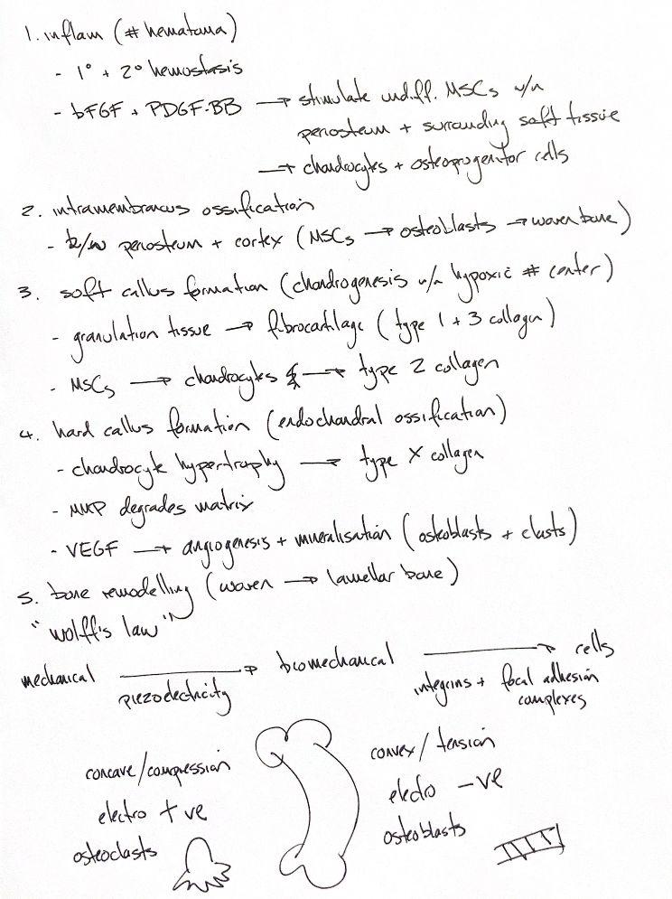
Read the draft explanation
Secondary healing has five overlapping phases.
1. Inflammation creates the fracture haematoma. Platelets activate in primary haemostasis, and fibrin forms in secondary haemostasis. bFGF and PDGF-BB activate mesenchymal stem cells in the periosteum and nearby soft tissues. These progenitors support cartilage and bone formation.
2. Intramembranous ossification starts between periosteum and cortex. Stem cells become osteoblasts and directly deposit woven bone. This early bone does not yet bridge the whole fracture.
3. Soft callus forms in the hypoxic centre. Granulation tissue becomes fibrovascular tissue and fibrocartilage, with types I and III collagen. Chondrogenesis then produces chondrocytes and a matrix rich in type II collagen.
4. Endochondral ossification creates hard callus. Chondrocytes enlarge and express type X collagen. Matrix metalloproteinases, or MMPs, break down matrix. VEGF supports vessel growth. Mineralisation and vascular invasion develop together. Osteoclasts remove mineralised cartilage; osteoblasts replace it with woven bone.
5. Remodelling replaces woven bone with lamellar bone. Wolff's law describes adaptation to loading. Integrins and focal adhesions help cells turn mechanical signals into biochemical responses: mechanotransduction.
For the electrical mnemonic, classic bending experiments found compression negative relative to other regions. Tension is shown positive in that simple model. This reverses the signs in the original notes and supplied textbook paragraph. Piezoelectric and fluid-flow signals are parts of a wider sensing system. Compression does not always mean osteoclasts, nor tension always osteoblasts; the loading pattern and biological setting determine formation and resorption.
References
Johnston & Tobias, 2nd ed., Ch. 39. Bassett & Becker 1962: https://doi.org/10.1126/science.137.3535.1063; Gross & Williams 1982: https://doi.org/10.1016/0021-9290(82)90174-9