
How secondary healing reduces strain
Read the draft explanation
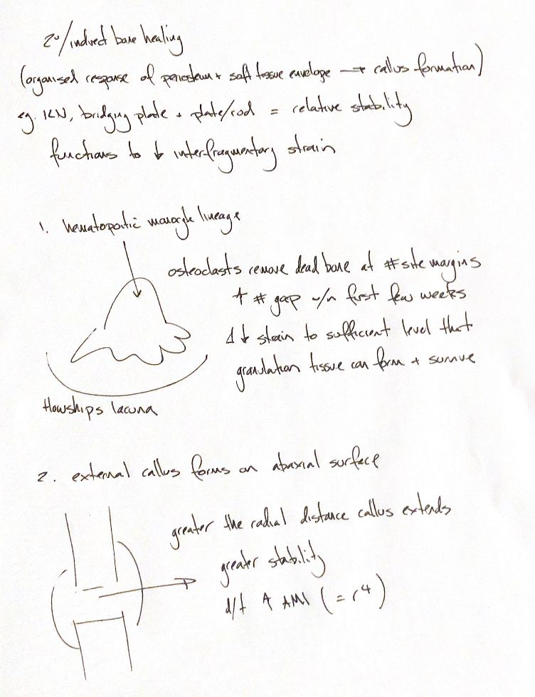
Secondary healing forms a callus through an organised response of the periosteum and surrounding soft tissues. Relative stability permits some movement. Interlocking nails, bridging plates and plate-rod constructs can provide this environment.
Two changes reduce interfragmentary strain as healing progresses.
First, osteoclasts remove dead bone from the fracture margins. These cells arise from the haematopoietic monocyte–macrophage lineage. Each cell resorbs bone within a Howship lacuna. The gap can therefore look wider on early radiographs, often during the first few weeks. For the same amount of movement, a wider gap has a smaller proportional change in length. Strain equals movement divided by the original gap length. As strain falls, granulation tissue can form and survive.
Second, callus grows around the outside of the fracture, on its abaxial surface. Its larger radius makes the repair more resistant to bending. As stability increases, movement at the gap falls.
The radius-to-the-fourth cue comes from the area moment of inertia of an ideal cylinder. For a solid circular section, I = πr⁴/4. A hollow tube depends on both its outer and inner radii. The point is geometric: moving material farther from the bending axis greatly increases stiffness. It is a fourth-power relationship, rather than a literal exponential law for every callus.
References
Johnston & Tobias, Veterinary Surgery: Small Animal, 2nd ed., Ch. 39, secondary healing and mechanics of bone.