
Primary healing - contact and gap
Read the draft explanation
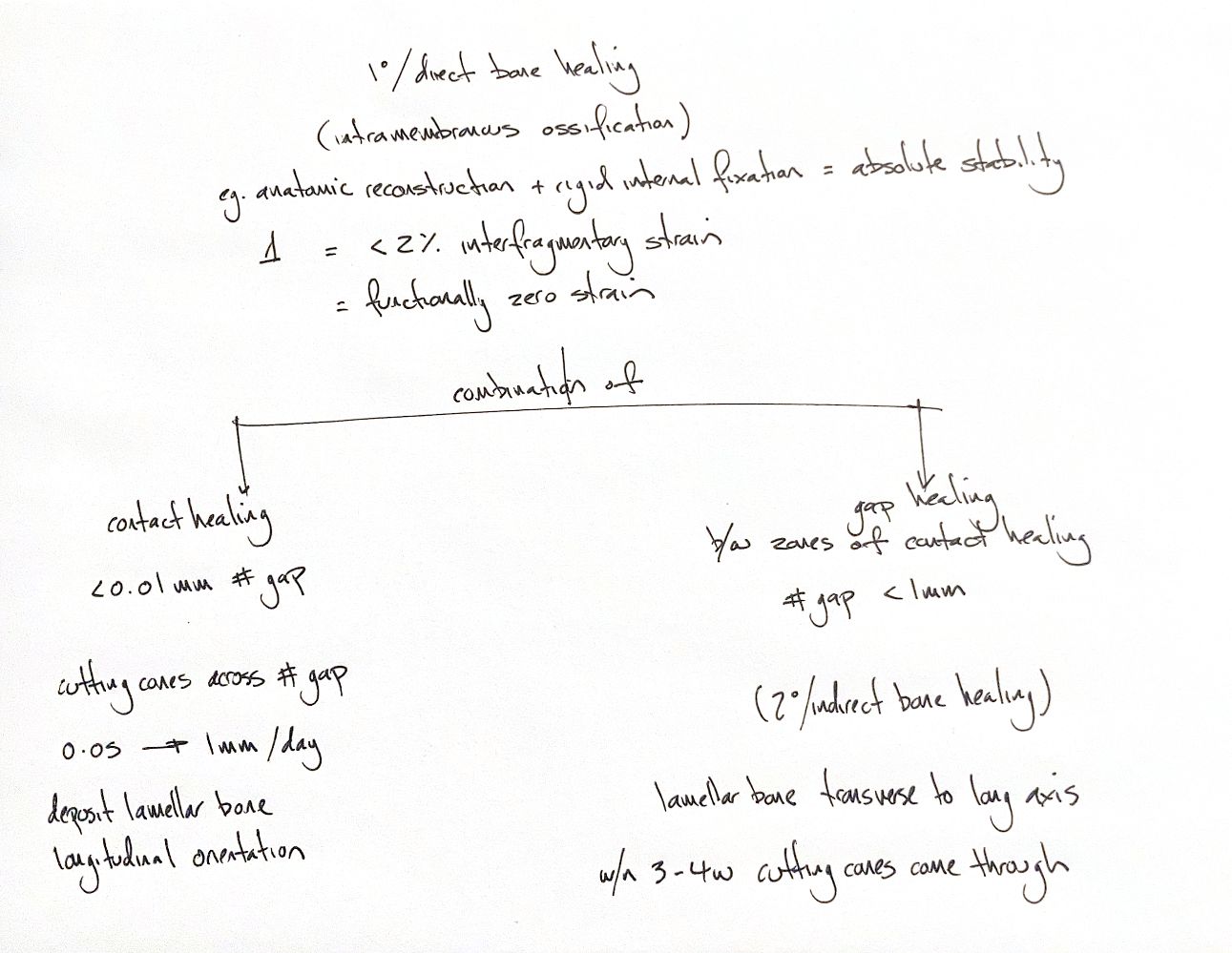
Primary healing is direct bone repair under absolute stability. It forms bone directly through intramembranous ossification. Accurate reduction, compression and rigid fixation make strain at the fracture functionally negligible. In the classic cortical-healing framework, interfragmentary strain is below 2%. Contact and gap healing occur together because the entire fracture surface never fits perfectly.
At contact sites, bone ends are less than 0.01 mm apart: 10 micrometres. Osteoclasts form the tip of a cutting cone. They remove damaged bone as they cross the fracture. A capillary and osteoblasts follow. Osteoblasts lay lamellar bone parallel to the long axis, joining the two cortices.
The reported cutting-cone rate is 50–100 micrometres per day, or 0.05–0.1 mm/day. The upper value is 0.1, not 1 mm/day.
Gap healing fills small spaces between stable contact zones. The gap is up to approximately 1 mm, with strain still below 2%. Fibrin, vascular sprouts and matrix fill the space. Osteoblasts deposit lamellar bone across it. These first lamellae lie transverse to the long axis, so the early union remains mechanically weak.
After about 3–4 weeks in the classic account, cutting cones cross from neighbouring and newly formed osteons. They replace the transverse scaffold with longitudinally aligned lamellar bone. These dimensions and timings describe the traditional cortical model; they are not universal cutoffs or a patient's healing deadline.
Gap healing is still primary healing. “Secondary osteonal reconstruction” describes its later remodelling step; it does not make gap healing secondary fracture healing.
References
Johnston & Tobias, Veterinary Surgery: Small Animal, 2nd ed., Ch. 39, contact and gap healing.