
Biological conditions for fracture healing
Read the draft explanation
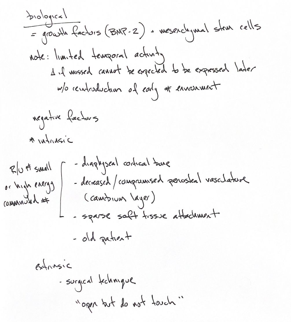
Fracture biology depends on both signals and living cells. Growth factors such as BMP-2 act alongside mesenchymal stem cells. The local environment guides these cells towards osteoblasts or chondroblasts, helping build bone or cartilage.
The periosteal cambium is an important cell source. The fracture haematoma, blood supply and attached soft tissues help support the early response. Growth factors act in an ordered sequence, and some signals have only a brief window of activity. If an early step is disrupted, later cellular responses may stall. Restoring an early fracture-healing environment may then be needed.
Some fractures start with less biological support. Diaphyseal cortical bone, poor periosteal blood supply, sparse soft tissue attachments and an older patient all reduce the available response. The cambium supplies progenitor cells; it should not be confused with the whole vascular periosteum.
Small-breed radius and ulna fractures are a useful example, especially distally, where soft tissue coverage and medullary vascular resources can be limited. A high-energy comminuted fracture creates a different problem: the injury may strip or devitalise periosteum, muscle and vascular attachments.
Surgical technique can add to that damage. The cue “open, but do not touch” means preserve fracture biology: limit unnecessary fragment handling and protect vessels, cambium and soft tissue attachments. It is a reminder to handle tissue carefully during the chosen approach.
References
hand-drawn ACVS Phase One notes; Johnston and Tobias, Veterinary Surgery: Small Animal, 2nd edition, Chapter 45; Welch et al., Vet Surg 1997. BMP-2’s requirement for initiation was demonstrated in a mouse model by Tsuji et al., Nat Genet 2006.