
Mechanical environment of fracture healing
Read the draft explanation
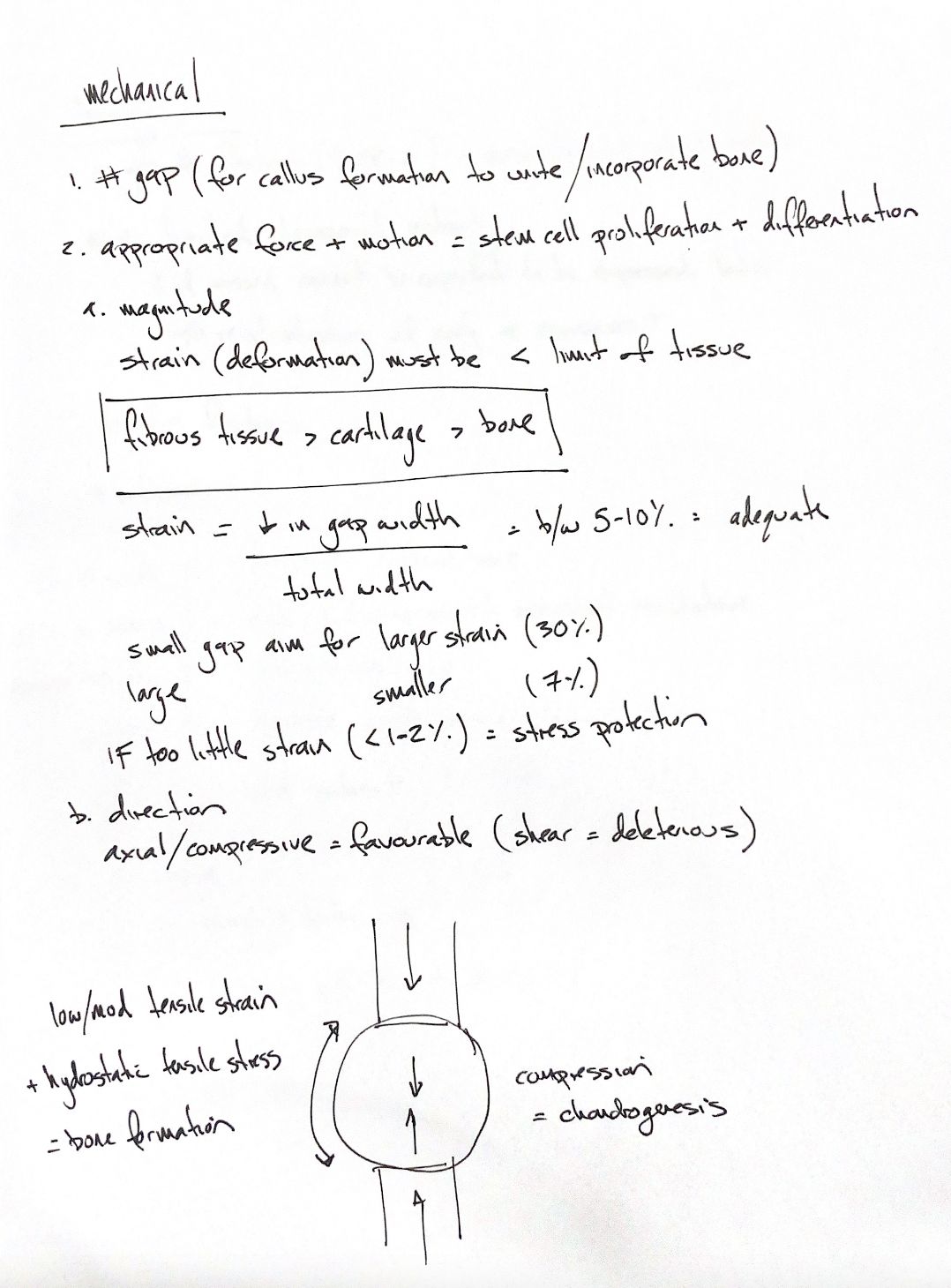
Fracture healing depends on both the gap geometry and the forces acting through it. Fragments must be close enough for the callus to unite them. Suitable motion can stimulate stem-cell proliferation and differentiation; too much deformation damages developing tissue.
The useful amount of motion depends on the model. In 42 sheep with externally fixed metatarsal osteotomies, Claes and colleagues compared 1, 2 and 6 mm gaps at about 7% or 31% initial interfragmentary strain. After nine weeks, higher strain produced more periosteal callus in the smaller gaps, but not in the 6 mm gaps. Larger gaps also reduced bending stiffness. The sketch’s 30% cue is a rough reference; this experiment tested about 31%. The response depended on gap size; neither value is a general clinical target.
For simple axial loading with gaps or comminution of a few to several millimetres, the textbook gives 5–10% as a guide. It also describes stress protection: very little mechanical stimulation may limit callus development. Its concern about strain below 1–2% belongs to that secondary-healing context. Direct healing under absolute stability deliberately uses very low movement. The two settings need different goals.
Direction matters too. In the callus sketch, axial compression makes the callus bulge. Cells near its centre experience compression, favouring cartilage formation. At the periphery, low to moderate tensile strain and hydrostatic tensile stress favour bone formation. Hydrostatic tensile stress describes the mean tensile part of the local stress state. These are local tissue signals; an axial force does not create the same strain everywhere.
Shear makes adjacent fragments slide past each other. It is less well tolerated than controlled axial movement and can delay healing. The aim is suitable stability for the intended healing pathway, while protecting the local blood supply and developing tissues.
References
Tobias & Johnston, Veterinary Surgery: Small Animal, 2nd ed., Chs. 39 and 45; Claes et al., J Orthop Res, 1997; Augat et al., J Orthop Res, 2003.