
Interfragmentary strain
Read the draft explanation
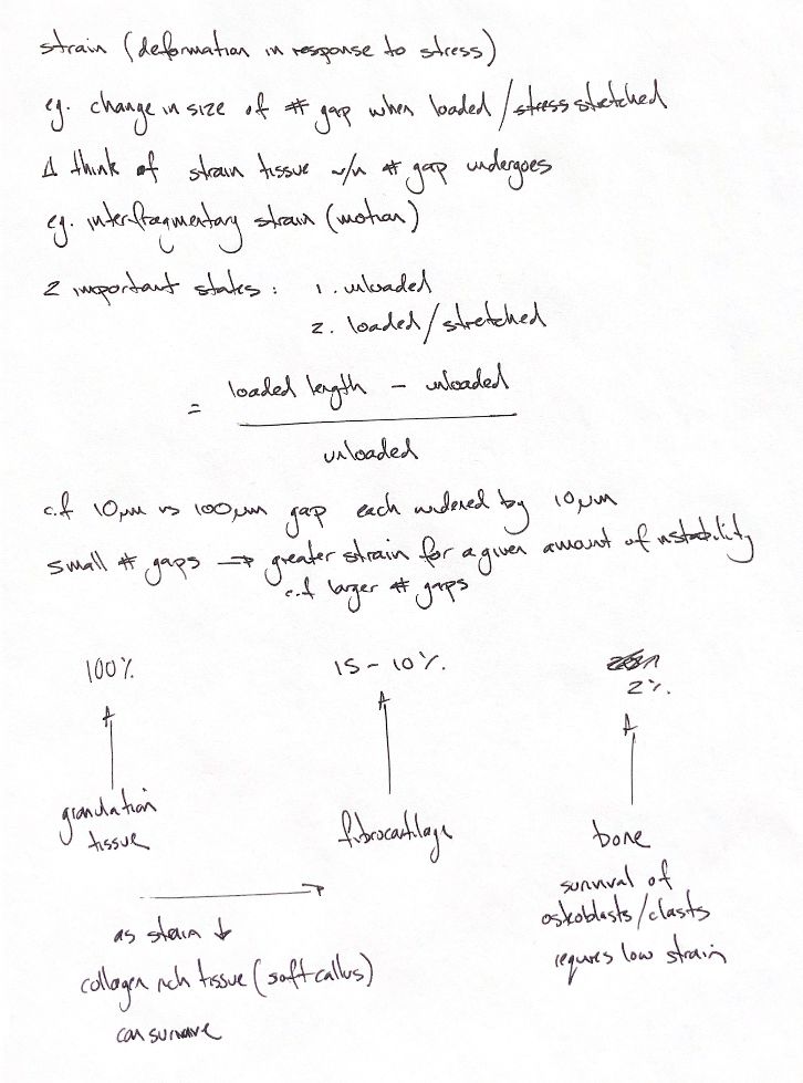
Interfragmentary strain describes movement relative to the starting fracture gap. First compare the unloaded and loaded states. Subtract the original gap width from the loaded width, then divide by the original width. Multiply by 100 to express it as a percentage. Taking the absolute change gives the magnitude, whether the gap opens or closes.
Now give two gaps the same movement. A 10-micrometre gap that widens by 10 micrometres has 100% strain. A 100-micrometre gap that widens by the same amount has 10% strain. The smaller gap has the greater strain potential. These are worked calculations, not measurements from patients.
Strain potential is a useful distinction. The calculation describes the change across a gap. It does not directly measure strain in every cell or part of the callus. Whole-construct displacement may also include implant bending and movement elsewhere. It cannot simply replace the change measured at the fracture gap.
The classic strain model links tissue tolerance to the tissues that can persist during healing. In this model, granulation tissue may tolerate almost 100% deformation, fibrocartilage about 10–15%, and bone about 2%. Granulation tissue is flexible early repair tissue. As motion falls, the collagen-rich soft callus and then bone can survive. Fibrous tissue tolerates more deformation than cartilage, and cartilage more than bone.
These are simplified tissue-tolerance estimates, not universal strain targets for a healing fracture. Bone formation needs a locally protected environment for osteoblasts and osteocytes. The loading direction, healing stage and local biology also matter. A larger gap may reduce the calculated strain for the same movement, but it also leaves more distance for tissue to bridge. Widening a gap is not a treatment goal.
References
Tobias & Johnston, Veterinary Surgery: Small Animal, 2nd ed., Chs. 39 and 45; Perren, Clin Orthop Relat Res, 1979.