
Two pathways and intramembranous ossification
Read the draft explanation
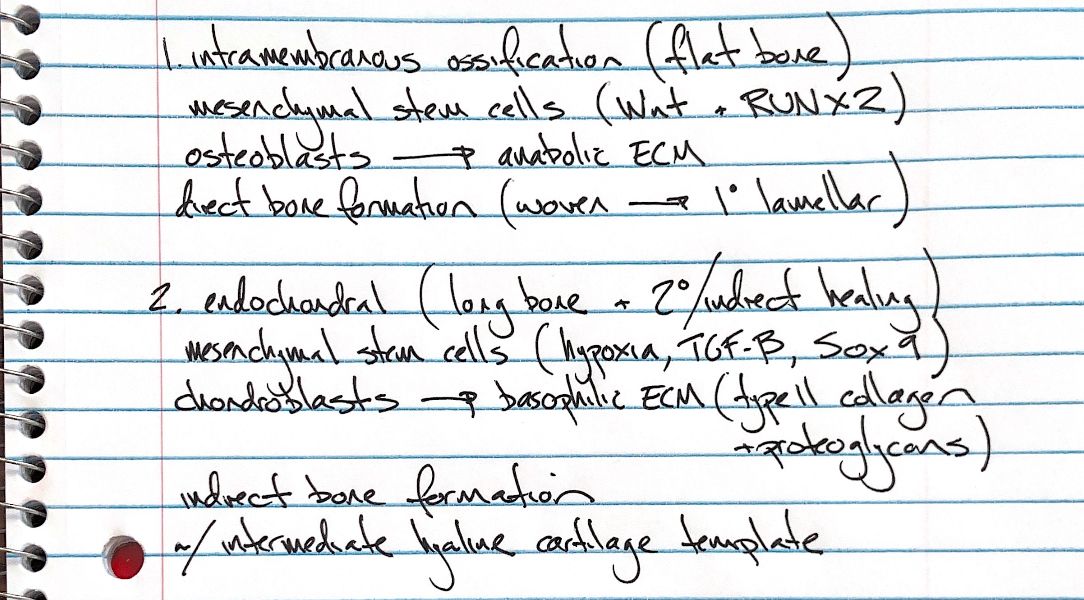
Bone can form directly, or it can replace a cartilage template. The starting cells are mesenchymal stem cells, but the cells that build the first matrix differ.
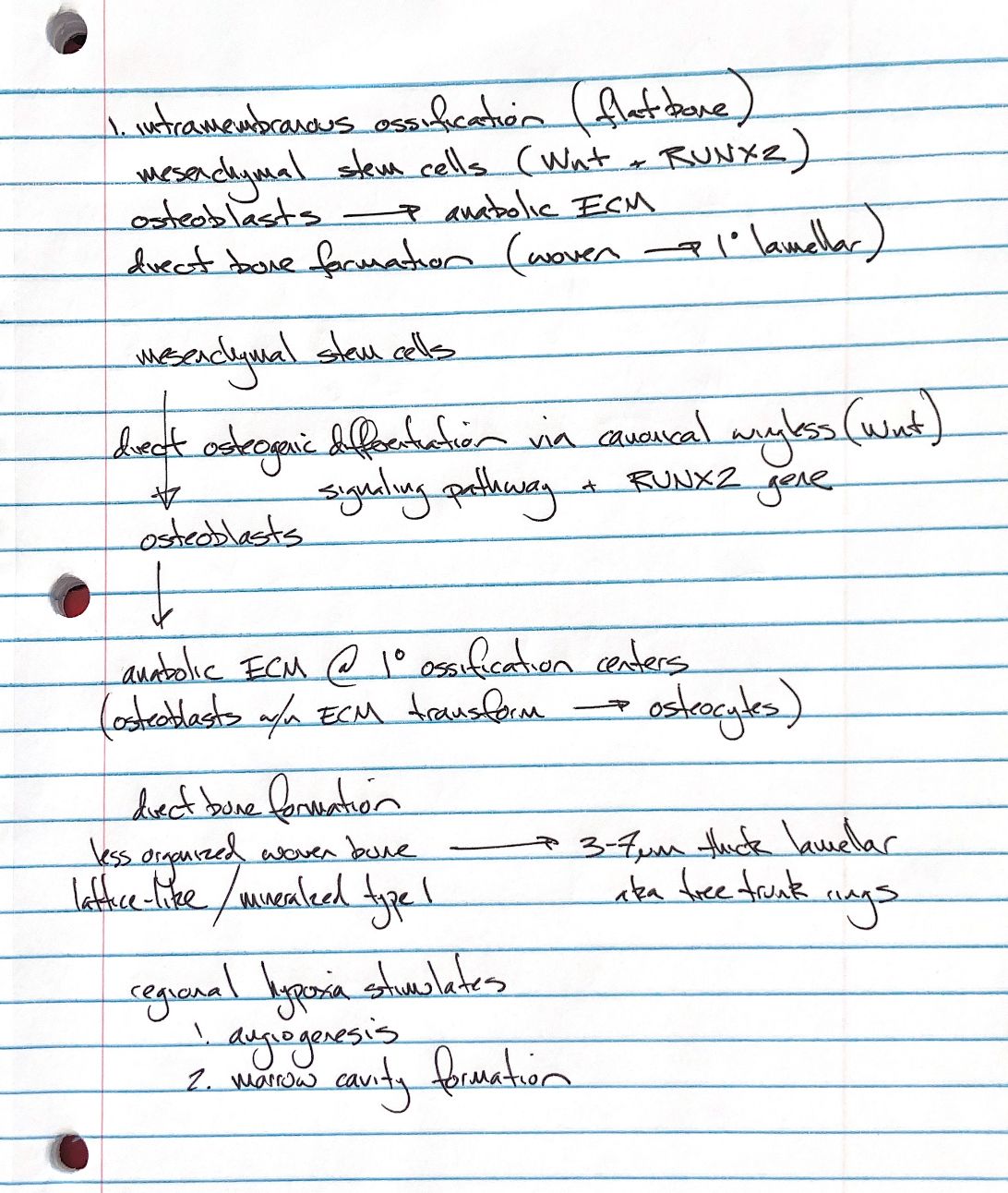
In intramembranous ossification, the cells become osteoblasts. Canonical Wnt signalling and the RUNX2 transcription factor help drive this change. Osteoblasts build extracellular matrix, or ECM, at primary ossification centres. As they become enclosed in that matrix, they become osteocytes.
Bone forms directly, without a cartilage stage. Early woven bone has a loose, irregular arrangement of mineralised type I collagen. It is then organised into primary lamellar bone. Lamellae are thin sheets. In an osteon, they form rings around a central canal, like tree-trunk rings. Lamellae are generally 3–7 micrometres thick, with variation by species and site.
As the developing bone enlarges, local hypoxia means parts have too little oxygen. This promotes angiogenesis: new blood vessels grow into the tissue. Some bones formed this way, such as the mandible, also develop marrow cavities. The separate ossification centres join as the vascular network develops.
Intramembranous ossification forms many flat bones, including bones of the skull and the scapula. Endochondral ossification forms most bones, including long bones. In that pathway, chondroblasts first build a basophilic matrix rich in type II collagen and proteoglycans. Basophilic means it takes up basic histological stains. This creates the hyaline cartilage template that bone later replaces.
In fracture repair, direct healing forms bone without a cartilage callus. Secondary healing uses both intramembranous bone formation and endochondral replacement of cartilage. The two pathways are not exclusive to one repair pattern.
References
Tobias & Johnston, Veterinary Surgery: Small Animal, 2nd ed., Ch. 39.