
Tendon healing, strength, repair configuration and loading
Read the draft explanation
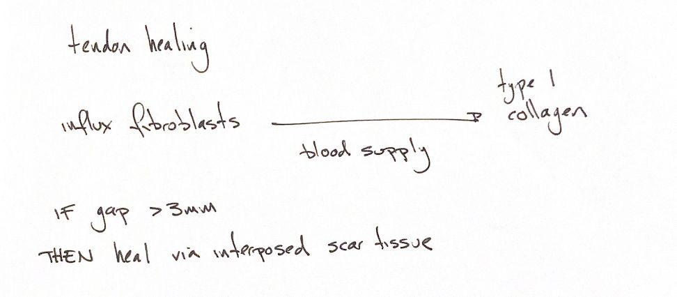
Tendon healing needs fibroblasts, blood supply and new collagen. As repair matures, type I collagen provides tensile support. A gap can fill with interposed scar. The 3 mm cue has a more specific meaning: in repaired canine intrasynovial flexors, gaps over 3 mm prevented the normal gain in strength and stiffness during early healing. It is not a universal scar threshold.
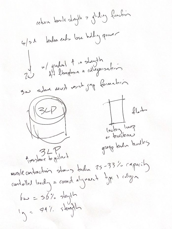
Early repair is vulnerable. Tendon ends lose holding power in the first 4–5 days, then gradually gain strength over the next two weeks with fibroplasia and collagen deposition. During the first three weeks, the suture construct remains central to resisting gapping and elongation.
The 56% and 79% figures come from a canine triceps-tenotomy experiment. Breaking strength was compared with the opposite unoperated muscle–tendon–bone unit. Four dogs were tested at six weeks and eight after at least a year. The reported ratios were 56% and 79%, respectively. At later testing, the repair site was no longer the weakest part of the unit. These are whole-unit experimental benchmarks for that model.
Match the holding pattern to tendon shape. A three-loop pulley suits round tendons and resists pull-out. Locking-loop and Krackow patterns suit flat tendons and grip collagen bundles. In many small-animal weight-bearing repairs, tensile strength takes priority over restoring glide.
Controlled loading helps collagen align. Loading from about 3–4 weeks is a textbook framework, with longer protection for some repairs. Timing remains specific to the repair. Its older 25–33% estimate compares muscle force with tendon strength; it does not mean 25–33% elongation strain or set an exercise dose.
References
hand-drawn ACVS Phase One notes; Johnston & Tobias, Veterinary Surgery: Small Animal, 2nd ed., Ch. 70. Dueland & Quenin, JAAHA 1980;16:507–512. Gelberman et al., JBJS Am 1999;81:975–982, https://pubmed.ncbi.nlm.nih.gov/10428129/.