
Muscle injury classification and the regeneration/scar balance
Read the draft explanation
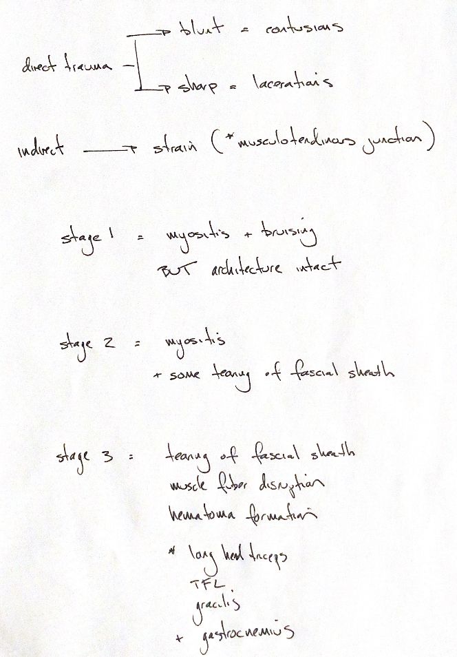
Blunt trauma causes a contusion. Sharp trauma causes a laceration. An indirect stretch injury causes a strain, most often at the musculotendinous junction.
Stage I means myositis and bruising with the muscle architecture intact. Stage II adds some tearing of the fascial sheath. Stage III disrupts the sheath and muscle fibres, with haematoma formation. These stages form a continuum. One injury can progress through them.
For stage III injuries, remember the long head of triceps brachii, tensor fasciae latae, gracilis and gastrocnemius.
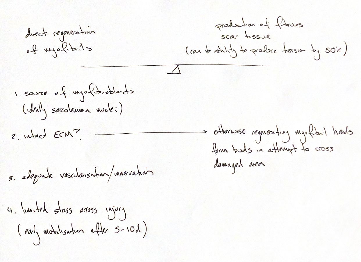
Muscle heals through both myofibril regeneration and fibrous scar formation. The balance matters. An intact endomysial scaffold guides repair. If the sheath is torn, regenerating ends send out buds to bridge the gap. Excess scar can block this growth, reduce function and leave the muscle prone to another injury.
Myoblasts build new muscle. Satellite cells supply these progenitors; mature myonuclei are not the cells that divide to make them. This is a myoblast source, rather than a source of scar-forming myofibroblasts. Preserving the local cell supply and sheath supports regeneration.
Repair also needs blood supply, innervation and limited stress across the injury. Once the initial injury has settled, controlled mobilisation can be considered after 5–10 days to help fibre alignment. Too much movement too soon can cause further damage.
Complete laceration can leave lasting force loss. In a rabbit extensor digitorum longus study, completely lacerated and repaired muscles recovered about 50% of normal tension after 12 weeks. Dense scar separated the muscle fragments. The figure reflects this injury and repair model.
References
Johnston & Tobias, Veterinary Surgery: Small Animal, 2nd ed., Chs. 38 and 70. Garrett et al. 1984: https://doi.org/10.1016/S0363-5023(84)80014-3. Dumont et al. 2015: https://doi.org/10.1002/cphy.c140068