
Tendon classes and coverings
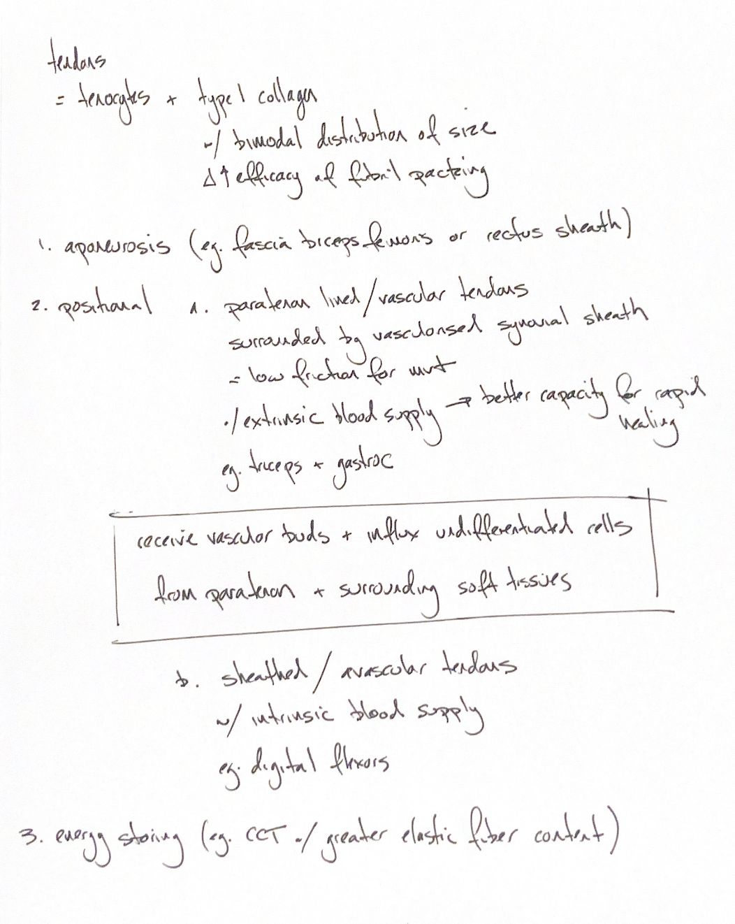
Read the draft explanation
Tendons carry force from muscle to the skeleton. Tenocytes sit among densely packed type I collagen fibres. Large and small collagen fibrils pack together efficiently.
The form matches the job. An aponeurosis is a broad, flat sheet, such as biceps femoris fascia or the rectus sheath. Positional tendons are relatively stiff and transfer muscle force to move a joint. Energy-storing tendons, including the common calcaneal tendon, have more elastic fibres. They store energy during loading and release it through recoil.
The covering also affects healing. Paratenon-lined tendons, such as the gastrocnemius and triceps insertion tendons, can receive new blood vessels and undifferentiated cells from the paratenon and nearby soft tissues. This extrinsic supply gives them a better capacity for rapid healing.
Sheathed tendons, including the digital flexors, depend more on their intrinsic blood supply for repair. These are often called “avascular” tendons, but the term is relative; they are not bloodless.
A paratenon and a synovial sheath are different coverings. The vascularised synovial sheath provides a low-friction tract for tendon movement. It should not be grouped as the paratenon itself.
References
hand-drawn ACVS Phase One study notes; Johnston and Tobias, Veterinary Surgery: Small Animal, 2nd edition, tendon structure and tendon-healing sections.