
Common calcaneal tendon
Read the draft explanation
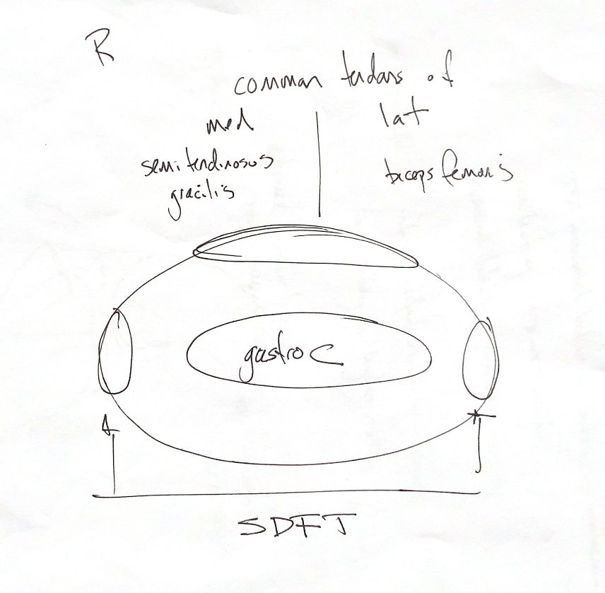
The common calcaneal tendon is a group of tendons, not a single strand. It brings together three main components: the gastrocnemius tendons, the superficial digital flexor tendon, and the combined tendon of biceps femoris, semitendinosus and gracilis.
For the right limb, keep the medial and lateral contributions straight. Semitendinosus and gracilis contribute medially. Biceps femoris contributes laterally. The central gastrocnemius group and the superficial digital flexor tendon complete the component map.
The map is a memory aid, rather than a fixed cross-section at every level. The superficial digital flexor tendon changes position as it travels distally and passes over the tuber calcanei. Its relationship to gastrocnemius therefore changes along the tendon.
References
hand-drawn ACVS Phase One study notes; Johnston and Tobias, Veterinary Surgery: Small Animal, 2nd edition, common calcanean tendon section. Canine component anatomy is also described by Lamb and Duvernois, Veterinary Radiology & Ultrasound, 2005;46:326–330, https://doi.org/10.1111/j.1740-8261.2005.00061.x.