
Muscle hierarchy, sarcomere and fibre types
Read the draft explanation
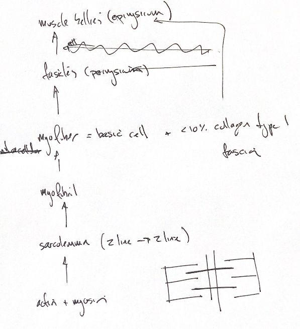
A muscle belly contains fascicles. Each fascicle contains myofibres, and each myofibre is one long cell packed with myofibrils. Repeating sarcomeres make up each myofibril. Actin and myosin form the overlapping filaments inside them.
Epimysium wraps the muscle belly, perimysium wraps each fascicle, and endomysium surrounds each fibre. These collagen-rich layers pass force towards the tendon. Type I collagen is abundant in this connective tissue. The less-than-10% estimate refers to muscle ECM, not to type I collagen alone.
Keep sarcomere and sarcolemma separate. A sarcomere runs from Z-line to Z-line. The sarcolemma is the fibre’s cell membrane.
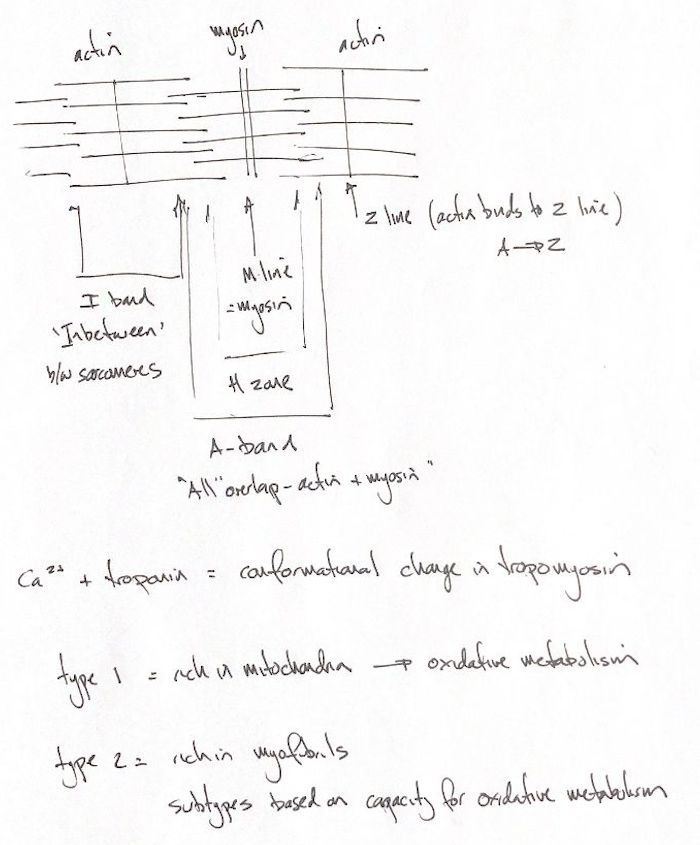
Actin attaches to the Z-line. The M-line lies in the centre. Titin links thick myosin filaments to the Z-line. The A-band spans the full thick-filament length, including the overlap zones. It is not just the overlap. The H-zone has thick filaments only. The I-band has thin filaments only and straddles the Z-line between adjacent sarcomeres.
A myosin molecule has a rod and two heads. Rods form the filament backbone; heads project into the bridge region. The vertebrate reference shows a 429 Å repeat, with about 143 Å between head crowns, and a 355 Å actin repeat. These are molecular repeat distances: 1 Å is 0.1 nm.
Calcium binds troponin C. Troponin I is inhibitory, and troponin T links the complex to tropomyosin. The complex changes shape, moving tropomyosin off actin’s myosin-binding sites. Myosin heads bind actin and use ATP hydrolysis to drive sliding. The sarcomere shortens as overlap increases.
Type I fibres have many mitochondria. Oxidative metabolism supports sustained, slower, lower-force work. Type II fibres have fewer mitochondria and more myofibrils. They produce faster, stronger, shorter contractions. Their subtypes differ in oxidative capacity, and the fibre mix varies within each muscle.
References
Johnston & Tobias, Veterinary Surgery: Small Animal, 2nd ed., Ch. 38, pp. 611–612. Squire 2019, Fig. 2: https://doi.org/10.3390/ijms20225715