
Connective-tissue matrix and mechanical roles
Read the draft explanation
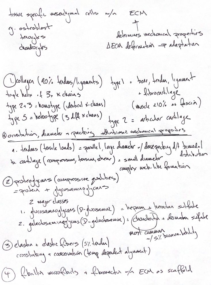
Connective tissue has tissue-specific mesenchymal cells within an extracellular matrix, or ECM. Osteoblasts, tenocytes and chondrocytes maintain different matrices. The matrix bears most of the load and sets much of the tissue’s mechanical behaviour. Cells sense its deformation and adapt its composition.
Collagen resists tension. It can make up to 90% of tendon or ligament dry weight. Each molecule has three alpha chains in a triple helix. Types II and III are homotypic: identical chains. Type V is heterotypic and can contain up to three different chains. Type I predominates in bone, tendon, ligament and fibrocartilage. Type II predominates in articular cartilage. ECM makes up less than 10% of skeletal muscle. Its type I-rich myofascia helps transfer force to tendon.
Orientation, diameter and packing shape collagen’s behaviour. Tendons use densely packed parallel fibres to resist tensile loads. Their fibrils have two main size groups: a bimodal distribution. Articular cartilage uses smaller fibrils in a web to resist deformation under tension, compression and shear.
Proteoglycans have a core protein with glycosaminoglycan, or GAG, side chains. Their charge attracts water and helps the matrix resist compression. Glucosaminoglycans include heparan and keratan sulfate, which contain D-glucosamine. Galactosaminoglycans include chondroitin and dermatan sulfate, which contain D-galactosamine. Chondroitin sulfate is the body’s most abundant GAG.
In eight male Beagles given a glucosamine and low-molecular-weight chondroitin combination, single oral dose bioavailability was 4.8–5.0%, measured as total chondroitin disaccharides. This does not measure intact chondroitin delivery to cartilage or clinical benefit.
Elastic fibres allow stretch and recoil. They form less than 5% of tendon dry weight. Tropoelastin aligns and assembles through temperature-dependent coacervation, then cross-links into elastin. Fibrillin microfibrils and fibronectin provide the scaffold for deposition.
References
Johnston & Tobias, Veterinary Surgery: Small Animal, 2nd ed., Ch38 and Ch68. Adebowale et al., 2002. https://pubmed.ncbi.nlm.nih.gov/12214321/