
Waveforms and coagulation
Read the draft explanation
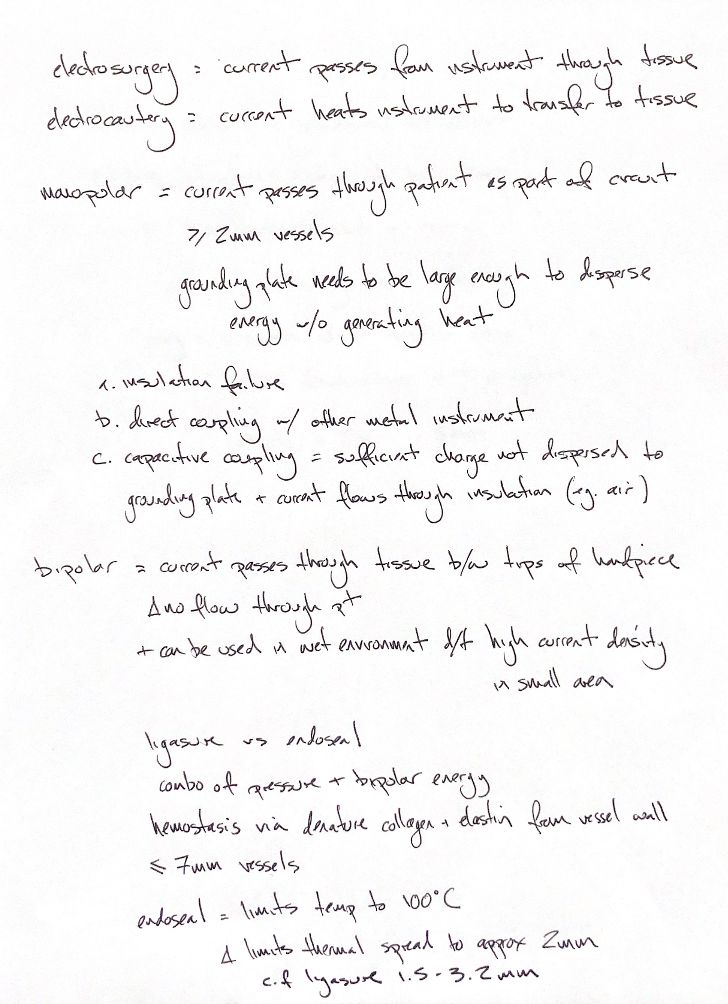
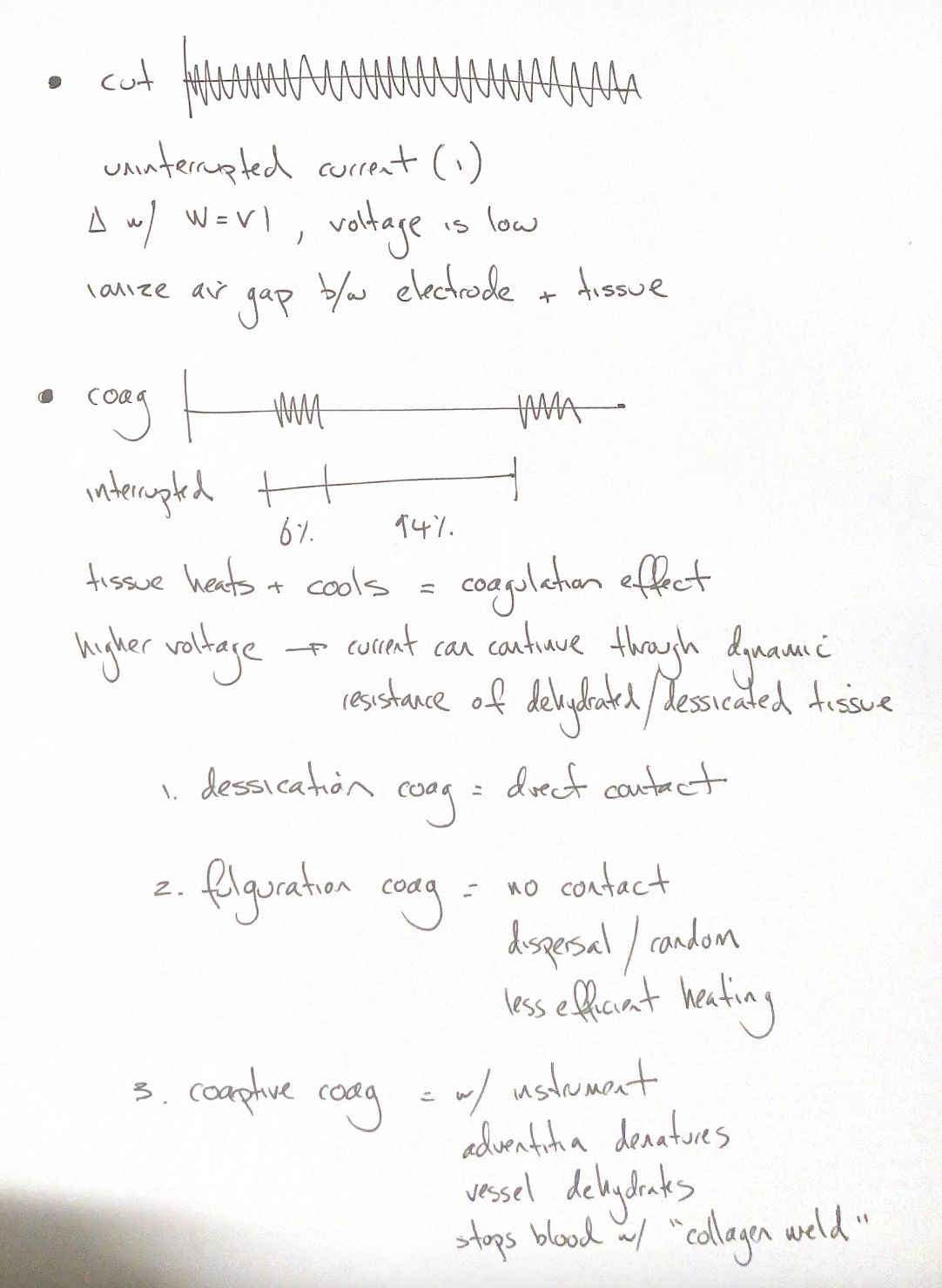
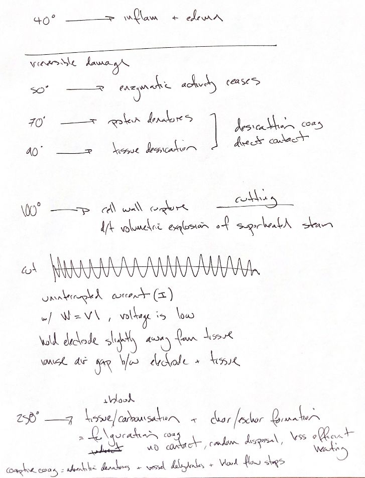
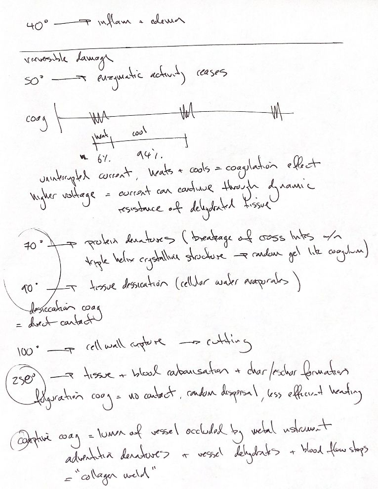
The waveform changes how heat reaches tissue. Pure cut delivers current continuously at lower peak voltage. The electrode sits just off the tissue. A focused arc crosses the ionised air gap and rapidly vaporises intracellular water.
A classic high-voltage coagulation waveform delivers short bursts. The illustrated example is on for 6% of the cycle and off for 94%. Tissue heats during each burst and cools between bursts. These are waveform examples, not fixed percentages for every generator or coagulation mode. Higher voltage lets current continue as drying tissue develops more impedance. Power follows W = V × I.
Contact changes the effect too. Desiccation uses direct contact. Water leaves the tissue and proteins denature into a coagulum. Fulguration holds the electrode off the surface. Random, dispersed sparks heat less efficiently than direct contact and create superficial carbonisation and eschar.
Coaptive coagulation adds compression. A metal instrument closes the vessel lumen before current is applied. Heat denatures the adventitia and dehydrates the vessel wall. The apposed walls form a collagen weld that stops flow.
References
Benitez, Johnston & Tobias, Veterinary Surgery: Small Animal,2nd ed., PDF pp.574–577; Brill, Obstet Gynecol Clin North Am 2011;38:687–702; Vilos & Rajakumar, J Minim Invasive Gynecol 2013;20:279–287.