
Temperature and tissue effect
Read the draft explanation
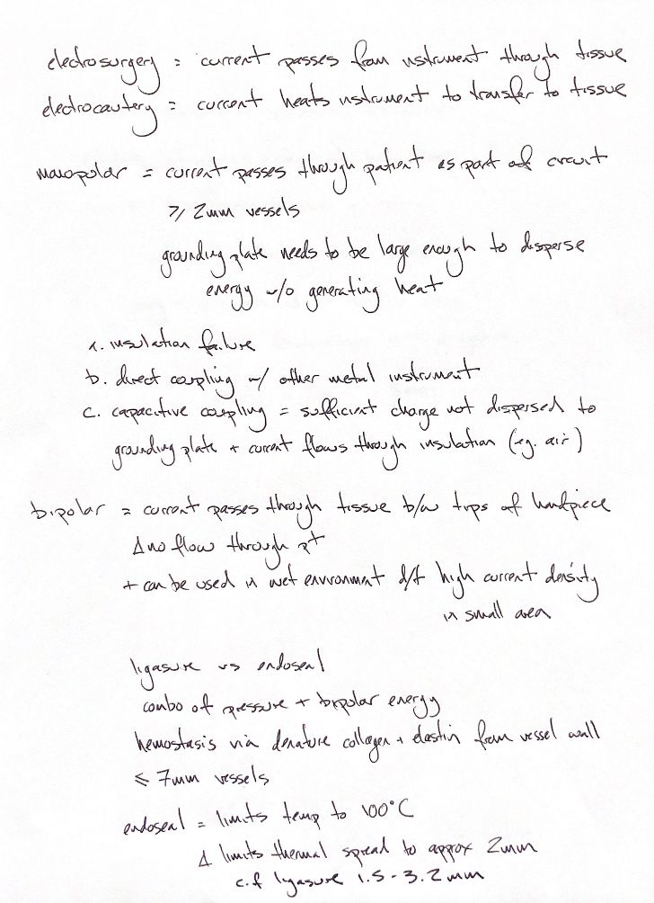
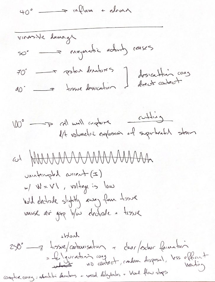
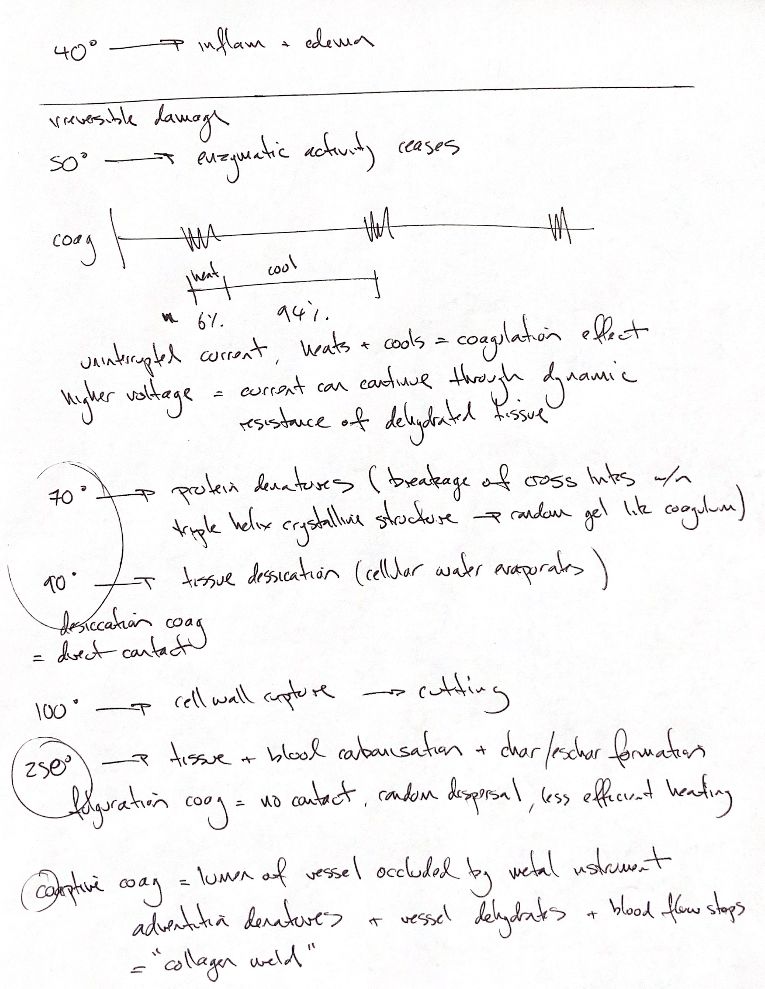
Temperature gives us a useful map of tissue effects, but exposure time matters. These are approximate landmarks, not instant switches.
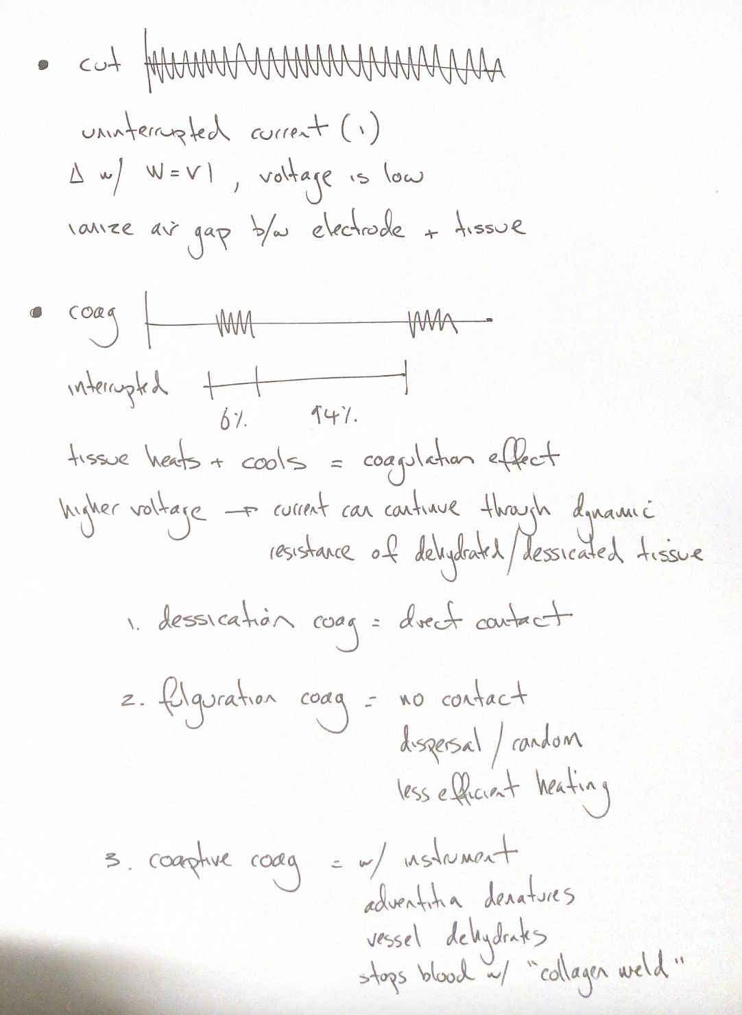
Around 40°C, tissue may develop inflammation and oedema. With sustained heating, enzymatic activity ceases around 50°C and injury becomes irreversible. By about 70°C, collagen cross-links break and the ordered triple helix becomes a gel-like coagulum. At about 90°C, cellular water evaporates and tissue desiccates. Direct-contact coagulation combines protein change with this water loss.
At about 100°C and above, intracellular water becomes superheated steam. Rapid expansion ruptures cells and cuts tissue. Animal cells have plasma membranes. Steam expansion ruptures the cell; there is no rigid cell wall.
At excessive temperatures, around 250°C in this teaching ladder, tissue and blood carbonise. Char and eschar form. A focused cutting arc concentrates energy into a narrow cleft. Fulguration spreads high-voltage sparks over the surface and favours carbonisation. Temperature, dwell time, current density and tissue impedance all shape the final effect.
References
Benitez, Johnston & Tobias, Veterinary Surgery: Small Animal,2nd ed., PDF pp.574–576, Table 15.1; Brill, Obstet Gynecol Clin North Am 2011;38:687–702; Dubiel et al., Vet Surg 2010;39:909–924.