
Margins - three-dimensional planning
Read the draft explanation
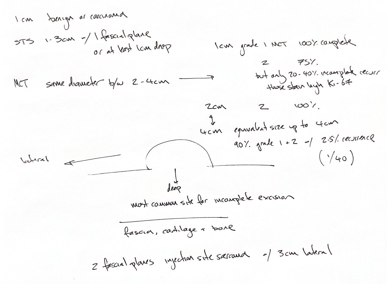
Plan a margin in three dimensions: around the tumour and beneath it. Tumour type, grade, size, site and surgical intent guide the plan. The 1-cm margins for selected benign lesions or carcinomas, and 1–3-cm canine STS margins, apply to specific settings. Fascia, cartilage and bone resist invasion. The deep margin is a common failure site.
Simpson prospectively mapped 23 cutaneous MCTs in 21 dogs. At 1 cm, margins were complete for all three Patnaik grade I tumours and 15/20 grade II tumours. At 2 cm, all grade II tumours were complete. Two grade II deep margins were complete but within 1 mm. These results apply to the grade I–II tumours studied, not grade III or larger tumours.
With proportional margins, the lateral margin equals the widest tumour diameter, up to 4 cm. Take at least one fascial plane deep. There is no automatic 2-cm minimum.
Pratschke's series included 40 dogs and 47 tumours. Of the 41 cutaneous tumours, 37 were Kiupel low grade: 90%. That is not a Patnaik I–II percentage. One suspected recurrence was unconfirmed. This is 1/47 tumours, about 2%, or 1/40 dogs, 2.5%. Neither is a confirmed recurrence rate.
Across series of incompletely excised grade II MCTs, recurrence was 18–35%. The broader 20–40% range is only a rough summary. Follow-up matters. Ki-67 with PCNA predicted local recurrence in Séguin's cohort. Low Ki-67 was linked to better survival in Ozaki's study.
For FISS, 3 cm is not a ceiling. Radical planning examples include 4–5-cm margins and two fascial planes. Imaging can show extension beyond what we can feel.
References
Johnston & Tobias, Veterinary Surgery: Small Animal, 2nd ed., chs25/82; Simpson2004 doi:10.2460/javma.2004.224.236; Pratschke2013 doi:10.2460/javma.243.10.1436; Séguin2006 PMID16955819; Ozaki2007 doi:10.1292/jvms.69.1117.