
Burn depth, zones and delayed healing
Read the draft explanation
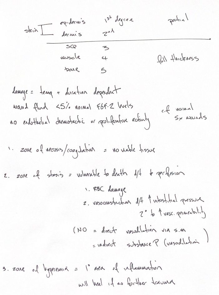
Burn depth depends on both the temperature and how long the tissue is exposed.
The five-degree scheme shown runs from epidermis to dermis, subcutis, muscle and fascia, then bone. Degrees 1–2 are grouped as partial thickness. Degree 3 and deeper are full thickness.
At the centre, the coagulation or necrosis zone has no viable tissue. Around it, the stasis zone still has threatened blood flow. Heat-damaged red cells become less flexible. Leaky capillaries raise pressure in the tissues, compress vessels and further reduce flow. The outer hyperaemic zone remains viable. It is inflamed but can heal if further injury is avoided.
Nitric oxide relaxes vascular smooth muscle directly. It can also promote release of substance P, which dilates vessels. This indirect pathway was supported by rat heat-exposure experiments, not a burn-treatment trial.
Burn fluid may also give a weaker early signal for new vessel growth. A small human study compared 7 surgical, 4 burn and 3 skin-graft donor samples. Mean FGF-2 was 914 ± 170 pg/mL in surgical fluid and 37 ± 7 pg/mL in burn fluid. The ratio is about 4%, which explains the <5% memory cue.
The tested burn-fluid samples caused no endothelial proliferation and no significant chemotactic activity. These findings suggest a weaker initial angiogenic stimulus than in deeper surgical wounds. They do not measure a fixed difference in healing time or establish a canine FGF-2 value.
References
Veterinary Surgery: Small Animal, Johnston & Tobias, 2nd ed., ch.81, redrawn from my study notes. Nissen et al., J Trauma 2003;54:1205–1210, DOI 10.1097/01.TA.0000061884.28845.5A, https://pubmed.ncbi.nlm.nih.gov/12813345/. NO mechanism: Yonehara & Yoshimura, Neurosci Lett 1999;271:199–201, DOI 10.1016/S0304-3940(99)00533-9.