
Assess, debride, then choose closure or open management
Read the draft explanation
Assess the tissue before deciding how to close a traumatic wound.
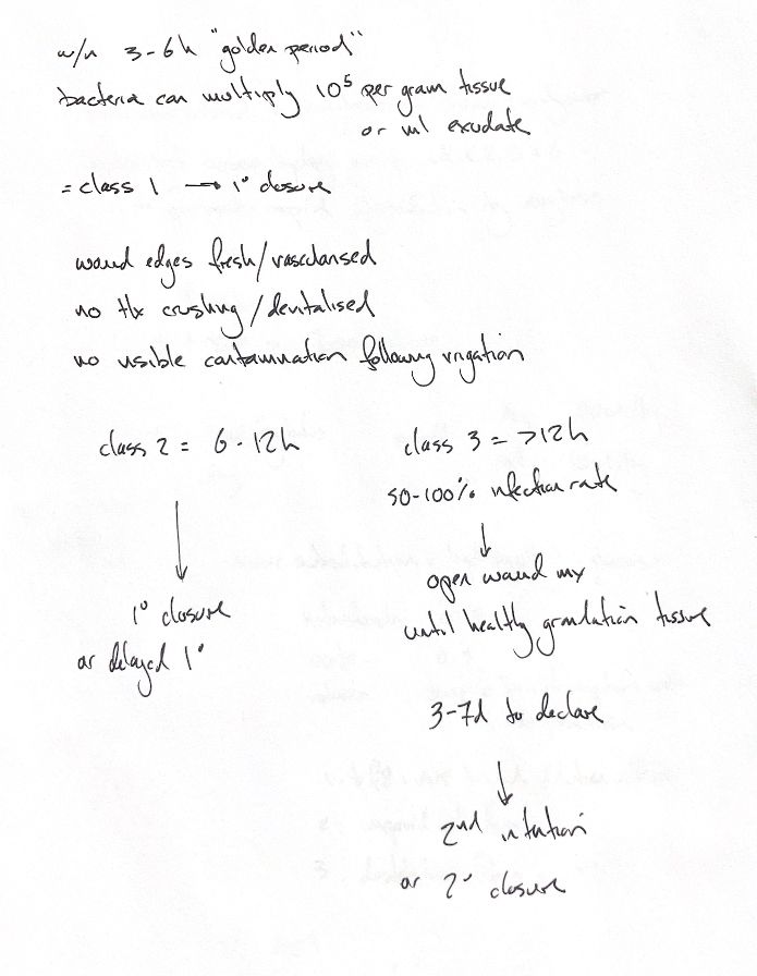
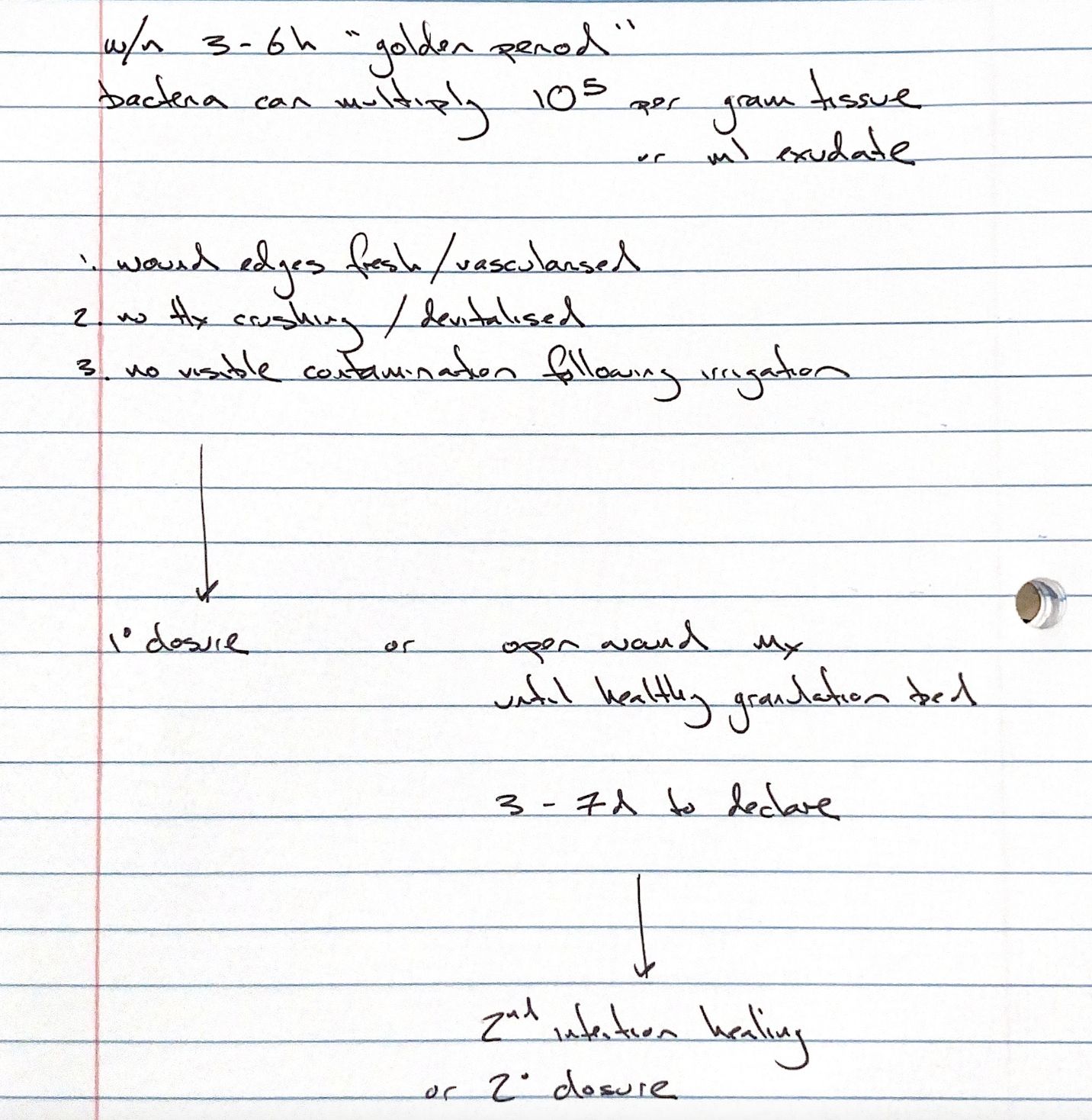
Primary closure may suit a healthy patient with fresh, well-perfused edges, no crush injury or dead tissue, and no visible contamination after irrigation. The traditional 3–6 h “golden period” helps frame that assessment. Tissue viability still takes priority.
Bacteria may multiply to 10⁵ organisms/g tissue or mL exudate during this period. Infection can also develop at lower counts, depending on the organism, host defences and local conditions.
If viability is unclear, contamination remains or the wound has dehisced, open management allows irrigation, debridement and repeated assessment. Crush necrosis may take 3–7 d to become clear; this does not tell us when granulation will form. Once a healthy granulation bed is present, we can consider secondary closure or let contraction and epithelialisation complete second-intention healing.
The age mnemonic uses “class I” for 3–6 h, “class II” for 6–12 h, with primary or delayed primary closure, and “class III” for >12 h, with open management. These are age-based study groups, separate from SSI classes. Age alone does not decide closure. The linked 50–100% infection figure refers to historical human guidance on closing contaminated wounds with foci of >10⁵ organisms/g, cited through Tobin (1984). It is not the infection rate for every veterinary wound over 12 h old.
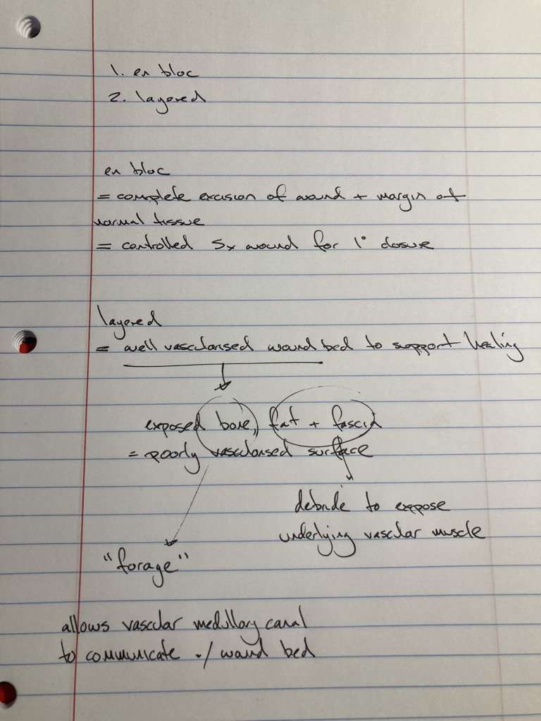
En bloc debridement removes the whole wound with a margin of normal tissue. This may create a controlled wound for primary closure. Layered debridement removes dead tissue step by step until a vascular bed is reached. Poorly vascularised fat or fascia may be removed to expose muscle. Forage of exposed cortex connects the vascular medullary canal to the wound bed. Preserve viable subcutis.
References
Johnston & Tobias, Veterinary Surgery: Small Animal, 2nd ed., chs 75–76; Steed et al., guideline 3.8: https://doi.org/10.1111/j.1524-475X.2006.00176.x