
Dogs and cats: closed incisions versus open wounds
Read the draft explanation
Dogs and cats heal differently. We also need to separate a closed incision from an open wound.
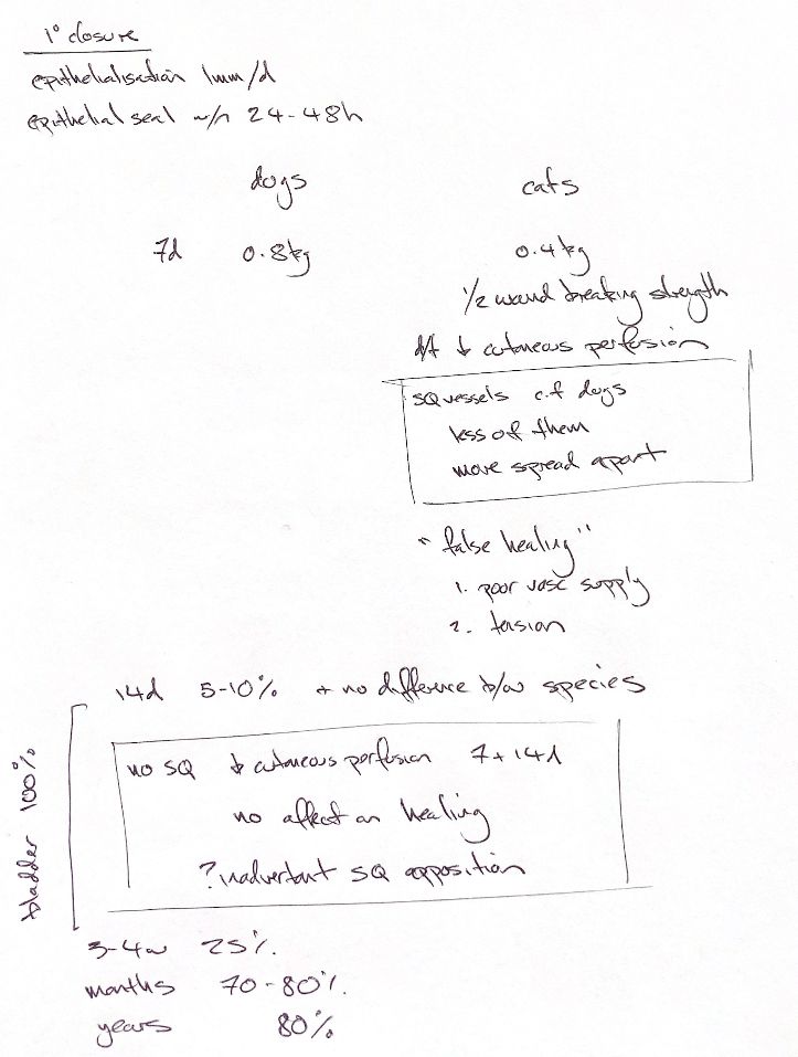
With ideal apposition, a closed incision epithelialises at about 1 mm/day and seals in 24–48 h. It still has little strength. Taking normal unincised skin as 100%, repair strength is about 5–10% at 14 d, 25% at 3–4 wk and 70–80% of ultimate strength after months. The figure of about 80% of normal at one year comes from rat incisions.
At day 7, experimental sutured incisions broke at 0.818 kg in dogs and 0.406 kg in cats; one dog strength specimen was excluded. Cats have lower early perfusion and fewer, more widely spaced trunk vessels. In open wounds, perfusion no longer differed by two weeks. This measured blood flow, not equal strength. Poor perfusion and tension contribute to feline problems, including “false healing”: the wound looks closed, then opens with normal motion after suture removal.
Removing subcutis reduced open-wound perfusion at days 7/14 in both species. It did not significantly change day-7 breaking strength within either species in sutured wounds. Apposition of the remaining subcutis was a proposed explanation, not a tested finding.
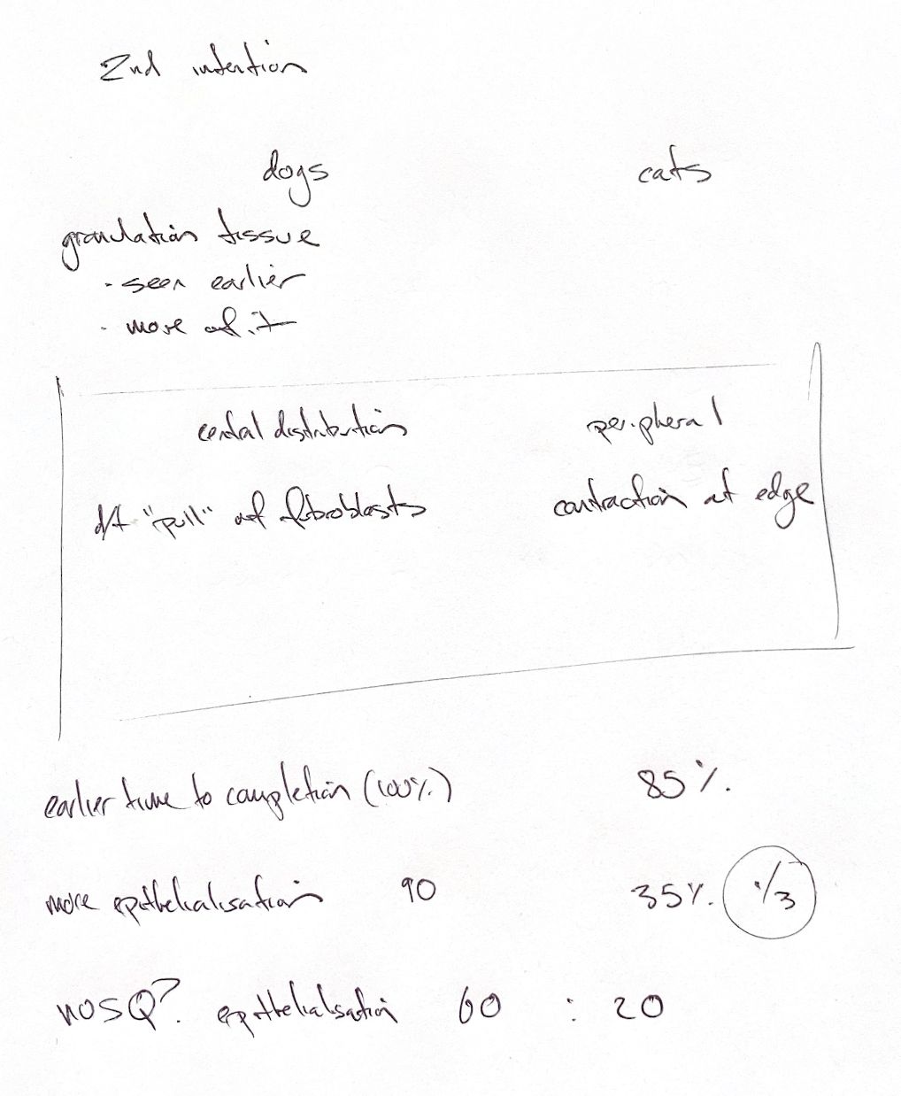
In open 2×2 cm dorsal wounds, dogs formed more granulation centrally; fibroblasts pulled inward and epithelium covered the bed. Cats formed less, mainly peripheral granulation, with more edge contraction. The 7.5 versus about 19 d figures describe complete granulation, not closure. Bottom coverage differed from filling the defect; one cat remained incomplete at 21 d.
At day 21, epithelialisation was about 89% in dogs and 34% in cats, roughly one-third. Total healing was about 98%/84%. Without subcutis, epithelialisation fell to about 61%/20%. Epithelialisation uses that day’s wound area. Total healing measures loss of open area from day 0, including contraction. Each experiment involved six dogs and six cats, so these are group results, not individual clinical forecasts.
References
Johnston & Tobias, Veterinary Surgery: Small Animal, 2nd ed., chs 9, 75–76; Bohling et al. 2004/2006; author study methods/tables: https://holocron.lib.auburn.edu/handle/10415/96