
Angiogenesis, fibroblasts and wound contraction
Read the draft explanation
New vessels, fibroblasts and contraction build the granulation-tissue bed together.
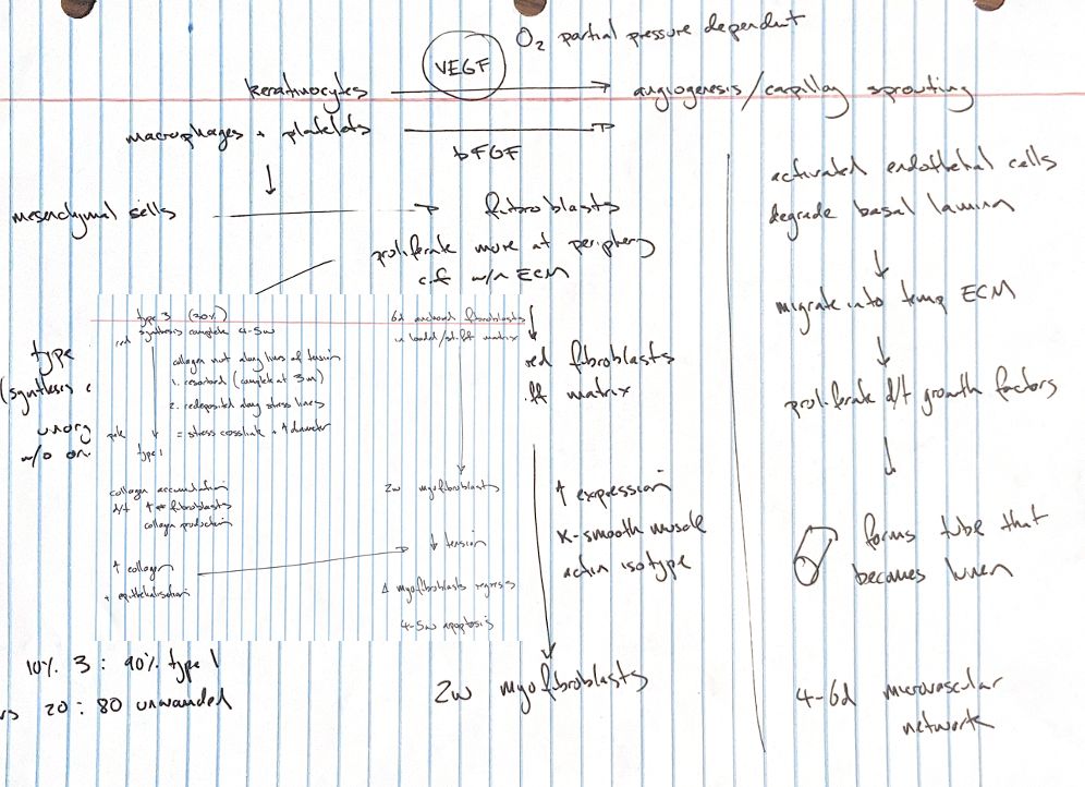
Inflammatory cells use oxygen while the injured blood supply delivers less. This creates local hypoxia. Wound lactate stimulates new vessel growth and collagen secretion. Disruption and hypoxia induce VEGF, mainly from wound-edge keratinocytes, but also from macrophages, endothelial cells, fibroblasts and platelets. VEGF and bFGF act in parallel.
Endothelial proteases open the parent vessel’s basal lamina. FGFs, PDGF, TGF-β and adhesion molecules help cells move into provisional extracellular matrix (ECM). VEGF and FGFs support cell division and formation of a tube with a lumen. A new basal lamina forms, followed by remodelling. Some vessels become arterioles or venules; others regress and are phagocytosed. The network brings oxygen and nutrients.
Like the neutrophil respiratory burst, endothelial responses need oxygen and depend on arterial PaO₂. Other regulators come from cells or matrix: HIF-1, TGF-α, EGF, TNF-α, nitric oxide, angiogenin, IL-8, lactate, fibronectin, fibrin and collagen. Their effects can promote or limit vessel growth, depending on context.
Mesenchymal-derived fibroblasts respond to macrophage and platelet signals. Collagen builds up according to cell number and output. Anchored fibroblasts divide less than cells arriving from the edges. TGF-β1 induces α-SMA and stress fibres, producing myofibroblasts. Attachments to ECM and nearby cells align with tension and transmit contraction. Stromelysin-1 and other MMPs also contribute.
The timing shown is approximate. TGF-β increases type I collagen, lowers MMP production and raises MMP inhibitors. Off-axis fibres are removed and collagen is laid down again. Aligned bundles cross-link and thicken. Type III:I is about 20:80 in unwounded dermis and 10:90 in mature scar. Collagen deposition and epithelial cover reduce tension, allowing myofibroblasts to regress and undergo apoptosis.
References
Johnston & Tobias, Veterinary Surgery: Small Animal, 2nd ed., ch. 9. Redrawn from my surgical study notes.