
Epithelialisation
Read the draft explanation
Epithelialisation starts at the wound edge within hours. It may take several days before we can see it.
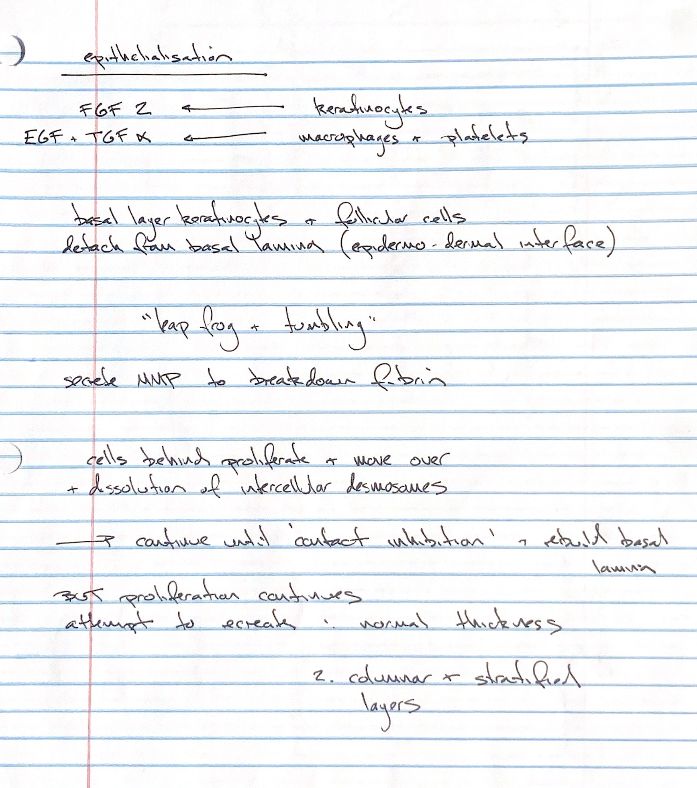
FGF-2 from keratinocytes, and EGF and TGF-α from activated platelets and macrophages, help release the cells. Basal keratinocytes and follicular epidermal cells detach from the basal lamina at the epidermo-dermal interface.
Desmosomes hold neighbouring cells together. Hemidesmosomes attach cells to the layer beneath them. As these attachments dissolve, the cells can move. Activated keratinocytes express integrins, which bind matrix proteins such as fibronectin. The cells advance by leapfrogging and tumbling.
Keratinocytes and endothelial cells release matrix metalloproteinases, or MMPs. These break down extracellular matrix, clearing the path ahead. Migrating keratinocytes also engulf debris by phagocytosis. Cells just behind the leading edge divide and advance over the wound bed.
When the advancing cells meet other epithelial cells, contact inhibition stops migration. The basal lamina is re-established. Cell division continues during remodelling and maturation as the epidermis attempts to regain its normal thickness, columnar organisation and stratified layers.
If the dermis is absent, its adnexal structures, including hair follicles, do not return. The newly epithelialised surface remains hairless.
References
Veterinary Surgery: Small Animal, Johnston & Tobias, 2nd ed., ch. 9. Redrawn from my surgical study notes.