Pre-referral oncology diagnostics
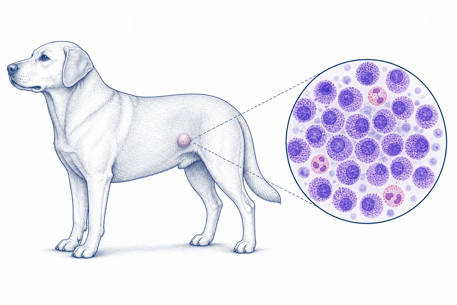 01Mast cell tumourA skin mass with mast cells identified on FNA
Clinical risk factors
Factors considered when planning surgery and the pricing guide.
- Maximum diameter and exact location
- Growth rate, ulceration, recurrence or regrowth
- Previous surgery or biopsy and the scar location
- Systemic signs and regional lymph-node findings
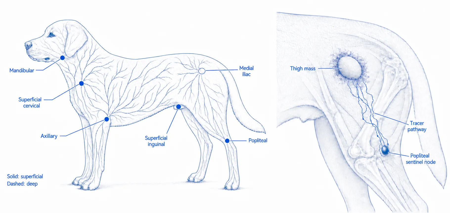
Sentinel-node mapping and risk-directed staging may change the operation
When to add abdominal or local staging
For uncomplicated cutaneous mast cell tumours, the routine staging priority is not the chest. Additional staging is selected when it could change the surgical plan.
Review the features that may change staging
- Large, fixed, ulcerated, rapidly growing, or recurrent masses
- Multiple mast cell tumours with systemic illness, an abnormal CBC, or a biopsy already showing high-grade disease
- Facial or muzzle, oral or perioral, preputial, or perineal location
Further staging may include abdominal ultrasound with targeted liver and spleen samples, local imaging for a deep or complex mass, or a preoperative biopsy when grade could change the surgical plan.
40%-63% may drain somewhere unexpected
Across published canine mast cell tumour studies, the mapped sentinel lymph node differed from the expected anatomical node in 40% to 63% of cases. This is why the mapped draining node, rather than only the nearest node, is important to surgical staging.
The closure and lymph-node field set the range
- Mass plus same-field lymph node
- $4,500-6,000
- Flap or graft reconstruction
- $5,800-8,000
- Separate-field lymph node
- Combined-procedure assessment
Pre-tax. A separate operative field requires its own setup and usually increases the primary package by one tier; a fixed Tier 4 price does not apply to every case. Staging is considered when combined work reaches Tier 4. Histopathology and routine rechecks for 12 months are included in these mass-removal packages. Review the full price guide.
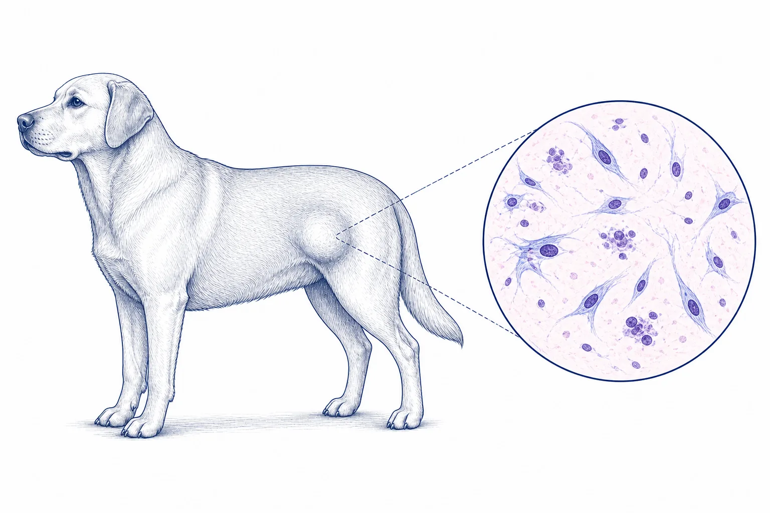 02Soft-tissue sarcomaA firm or deep mass with spindle or mesenchymal cells, or a nondiagnostic FNA that remains concerning
Clinical risk factors
Factors considered when planning surgery and the pricing guide.
- Maximum diameter in three dimensions, including whether it is over 5 cm
- Whether the mass is superficial or deep to the fascia, and mobile or fixed
- Whether it is recurrent or was previously removed without planned margins
- Any previous histopathology, including the reported grade, and the scar or biopsy-tract location
CT and biopsy are selected only when they could change the margins or closure plan
When contrast CT may be recommended
Contrast-enhanced CT is Dr Wood's usual additional local planning test when a mass is over 5 cm, deep, fixed, poorly defined, recurrent, close to a joint or an important nerve or blood vessel, or when wide margins in that location may require a flap, graft, or other closure plan.
CT shows the relationship between the mass and the tissues that may need to be removed or reconstructed. It does not provide a tumour grade.
When a preoperative biopsy may be recommended
A deliberately placed core or incisional biopsy may be useful for a distal-limb or other constrained mass when the result could change the margins, reconstruction, or whether to proceed with an operation that has substantial functional impact. The biopsy tract must be positioned so it can be removed with the definitive surgery.
Biopsy grade is not definitive. In a study of 68 dogs, the preoperative biopsy grade matched the complete-excision grade in 59% of soft-tissue sarcomas. It underestimated the final grade in 29% and overestimated it in 12%.
68-dog biopsy-grade study · spindle-cell sarcoma cytology study
The closure plan is the main price distinction
- Primary closure
- $3,000-4,700
- Flap or graft reconstruction
- $5,800-8,000
Pre-tax. Histopathology and routine rechecks for 12 months are included. Imaging or biopsy performed before definitive surgery is estimated separately. Review the full price guide.
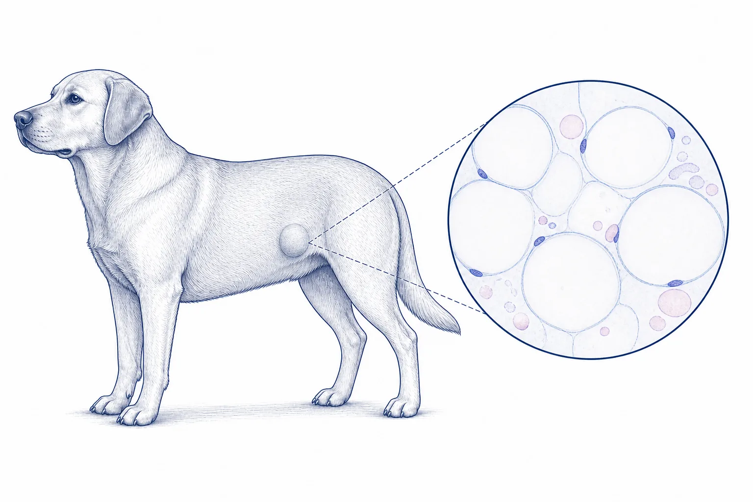 03LipomaAdipose tissue on FNA with a compatible superficial mass
Features that change planning
These findings help distinguish a simple superficial mass from one that needs CT planning.
- Maximum diameter, exact location, and whether the mass is superficial or deep
- Whether it is freely mobile, fixed, poorly defined, or within a muscle
- Any pain, altered movement, weakness, or other signs that may indicate nerve involvement
- Previous surgery and whether the mass has recurred
CT may be recommended for a deep or anatomically complex lipoma
Why CT and 3D planning may be useful
For a large, deep, intramuscular, recurrent, fixed, or anatomically complex lipoma, CT can map the muscle compartments, vessels, and the expected or visible course of important nerves. Dr Wood reviews the CT and performs his own 3D reconstruction for surgical planning.
CT can also suggest an infiltrative pattern. In a study of 60 adipose masses, 75% of infiltrative lipomas were irregular and 100% contained linear soft-tissue components. Histopathology provides the final diagnosis.
60-lesion CT comparison study · 11-dog intermuscular lipoma study
Simple mass removal
- Simple mass removal
- $3,000-4,700
Pre-tax. Histopathology and routine rechecks for 12 months are included. CT, when recommended, is planned separately. Review the full price guide.
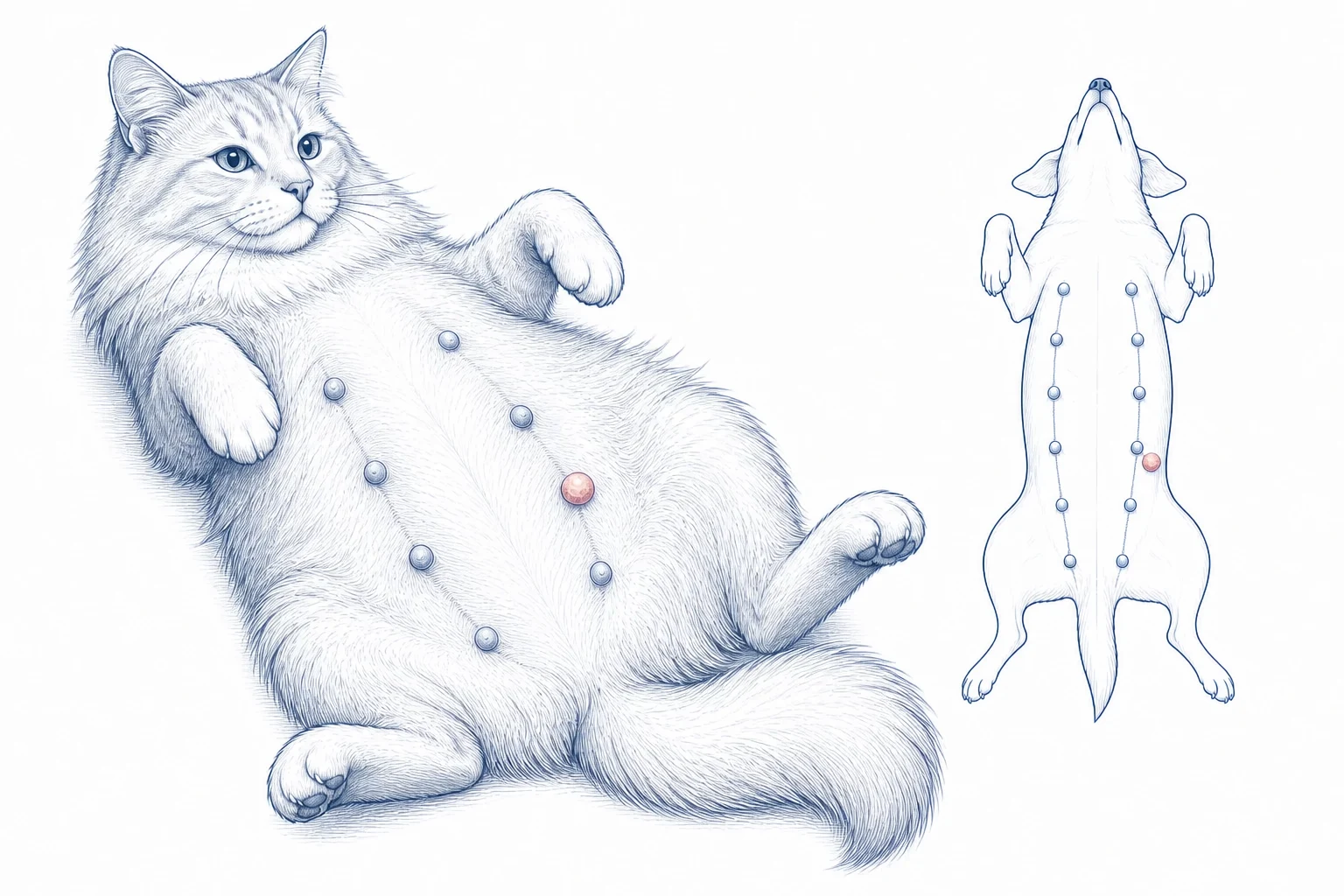 04Mammary massOne or more mammary lesions in a cat or dog
Clinical risk factors
Factors considered when planning surgery and the pricing guide.
- Affected gland and maximum diameter of every mass
- Growth rate, fixation, ulceration, discharge, or pain
- Axillary and superficial inguinal lymph-node findings
- Previous surgery or biopsy and the scar location
Species, distribution, staging, and closure needs determine the operation
Species and tumour distribution change the operation
Cats
Feline mammary tumours are more often malignant. When both chains require surgery, Dr Wood typically recommends staged bilateral mammary-chain surgery. The timing is planned from disease distribution, patient health, and closure requirements.
Dogs
The number, size, location, fixation, and lymphatic field determine whether a limited removal, regional procedure, or chain procedure is discussed.
Routine abdominal imaging is not usually required. The relevant regional lymph node can be assessed and sampled during the mammary procedure when indicated.
3 cm changes the prognosis discussion
Maximum tumour diameter is one of the simplest useful risk features to record before referral. It does not replace histology, but it helps frame staging and surgical urgency.
canine mammary tumour size study · feline mammary carcinoma study
Limited, unilateral, staged, and single-session bilateral surgery
- Limited removal with primary closure
- $3,000-4,700
- Feline unilateral mammary chain
- $4,500-6,000
- Staged second feline chain
- $3,000-4,700
- Feline bilateral chains, single session
- $7,800-10,500
Pre-tax. Histopathology and routine rechecks for 12 months are included. The examination determines whether limited removal, a unilateral procedure, staged treatment, or selected single-session bilateral surgery is appropriate. Review the full price guide.
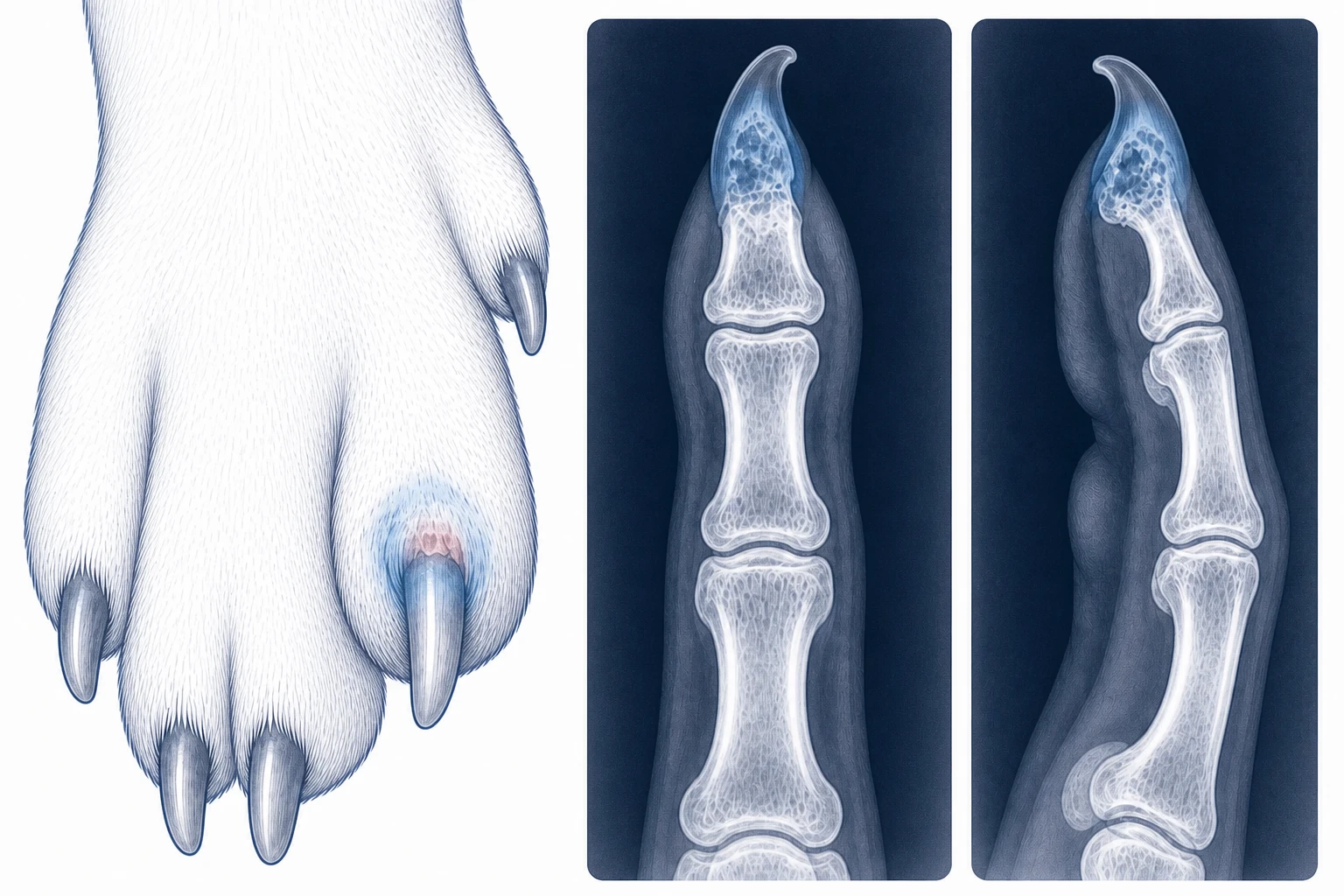 05Digit or nail-bed lesionPersistent nail loss, swelling, ulceration, pain, or bone lysis
Clinical risk factors
Factors considered when planning surgery and the pricing guide.
- Exact digit and maximum lesion diameter
- Nail loss or deviation, ulceration, discharge, or bleeding
- Duration, growth, pain, lameness, and response to treatment
- Previous surgery or biopsy and regional lymph-node findings
Imaging, tissue diagnosis, node assessment, and amputation planning are considered together
Match the operation to the species and imaging
Dogs
Persistent nail-bed lesions, especially with bone lysis, require a tumour work-up even when infection is also present. Radiographs and tissue diagnosis guide biopsy, digit amputation, and regional-node assessment.
Cats
A destructive digital lesion may be a primary tumour or metastatic disease. Thoracic imaging is particularly important because lung tumours can spread to the digits.
83% were malignant
In a 117-case study of canine digit masses with bone lysis, 83% were malignant. Infection can still coexist, so drainage or inflammation should not end the tumour work-up.
Digit amputation is usually a primary-closure procedure
- Digit amputation
- $3,000-4,700
- With same-field lymph node
- $4,500-6,000
Pre-tax. Histopathology and routine rechecks for 12 months are included. Staging and any preoperative biopsy or advanced imaging are planned separately. Review the full price guide.
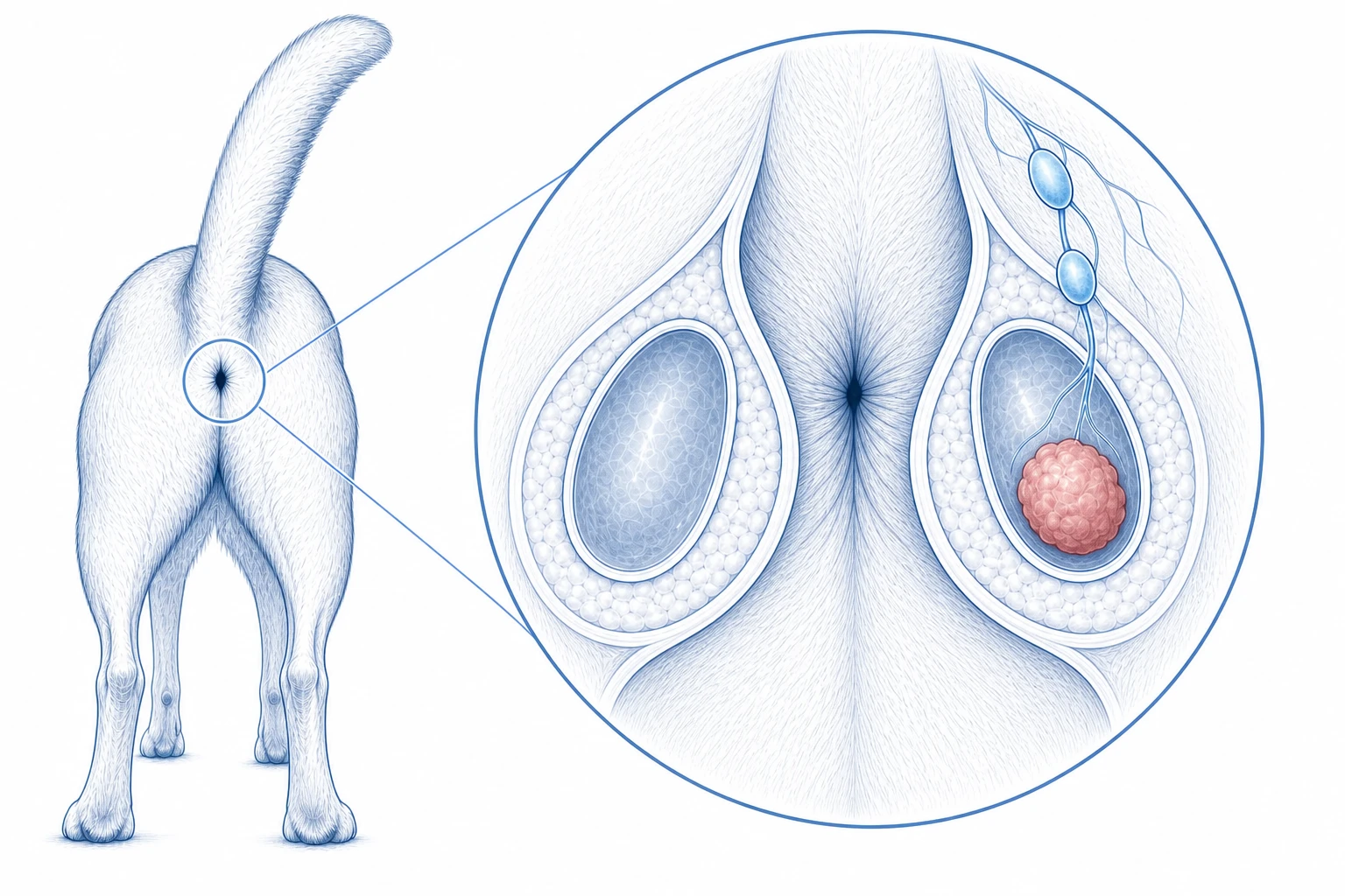 06Anal sac mass / suspected AGASACAAnal sac apocrine gland adenocarcinoma or another anal sac lesion
Clinical risk factors
Factors considered when planning surgery and the pricing guide.
- Affected side and maximum diameter, including whether it is over 2.5 cm
- Findings in the opposite anal sac and on rectal examination
- Tenesmus, constipation, stool change, thirst, urination, weakness, or appetite change
- Previous surgery or biopsy and available calcium results
CT, regional-node surgery, and bilateral anal-sac removal may be discussed
Use size as a risk marker, not a rule that replaces staging
The historical clinical scheme separates primary tumours at 2.5 cm. However, a small primary mass or normal calcium does not rule out regional nodal disease.
How CT findings are interpreted
- In one series, 20% of dogs with a primary mass under 2 cm already had confirmed lymph-node metastasis.
- One bilateral-surgery study treated a regional node over 5 mm, or abnormal in shape or internal appearance, as suspicious. Size alone does not confirm metastasis.
- Cytology or histopathology is used to establish nodal metastasis. Once nodal disease is confirmed, the published clinical scheme separates nodal stages at 4.5 cm.
- CT also shows the relationship to the rectum, pelvic canal, vessels, and ureters when surgery is planned.
small-primary metastasis study · bilateral anal-sac study and staging scheme
20% had hidden bilateral disease
A 2024 study found cancer in both anal sacs in 7 of 35 dogs whose preoperative work-up suggested disease on only one side. This supports discussing bilateral removal, while the final recommendation remains patient specific.
bilateral anal-sac study · CT versus ultrasound study · CT staging study
Unilateral and bilateral anal-sac removal
- One anal sac — Tier 2
- $4,500-6,000
- Both anal sacs — Tier 3
- $5,800-8,000
Pre-tax. See the named procedure entries for included care. Advanced imaging or a separate lymph-node operation is planned and estimated individually. Review the full price guide.
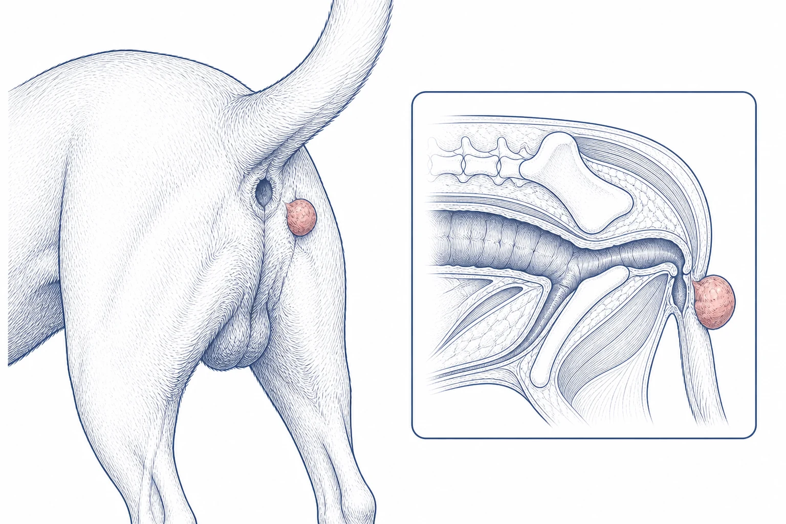 07Perineal massPerianal or perineal skin and gland presentation
Clinical risk factors
Factors considered when planning surgery and the pricing guide.
- Maximum diameter, including whether it is over 5 cm
- Fixation or deep invasion, ulceration, recurrence, or rapid growth
- Rectal examination, both anal sacs, and regional lymph-node findings
- Previous surgery or biopsy and the scar location
Most superficial masses proceed to a simple-removal discussion
CT or biopsy is selective, not routine
A small, superficial perianal-gland mass in a typical patient may need only FNA, rectal examination, and a simple-removal plan.
When CT or biopsy may be added
They may be recommended when a suspected perianal or hepatoid-gland tumour is over 5 cm, fixed or invasive, recurrent, of uncertain tissue origin, or when the result could change the margins or closure plan. The 5 cm finding does not apply to anal-sac cancer.
5 cm changes the risk discussion
For perianal or hepatoid-gland adenocarcinoma, published data associate tumours over 5 cm with a substantially higher risk of tumour-related death. Regional lymph-node spread has been reported in about 15% of dogs.
This evidence does not apply to anal-sac apocrine gland adenocarcinoma, which has a separate pathway above. Review the hepatoid-gland tumour evidence.
Most superficial perineal masses use the simple-removal tier
- Simple mass removal
- $3,000-4,700
- Flap or larger reconstruction, if required
- $5,800-8,000
Pre-tax. Histopathology and routine rechecks for 12 months are included. Neutering can be added to the surgical plan and estimate when recommended. A rectal, anal-sac, deep pelvic, or separate lymph-node procedure is estimated according to the operative fields involved. Review the full price guide.