
Thoracic lymphatic drainage
Read the draft explanation
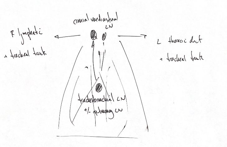
My sketch highlights the bronchial and mediastinal lymph-node groups. The patient's right is on the viewer's left. The patient's left is on the viewer's right.
Pulmonary lymph nodes receive lymph from the lungs and drain toward tracheobronchial nodes. Pulmonary nodes are not present in all dogs; that is the reason for the ± cue. Tracheobronchial nodes can drain into mediastinal nodes or other tracheobronchial nodes. They also receive lymph from structures beyond the lungs and bronchi.
Cranial mediastinal nodes vary in number and shape. The two upper symbols in my drawing are a schematic, not a fixed count for every animal.
On the right, mediastinal lymph can enter the right lymphatic duct, the right tracheal trunk, or both. On the left, it can enter the thoracic duct, the left tracheal trunk, or both. The + signs in the original group the possible outlets; they do not mean that every node must drain through both routes.
Canine lymphangiography has shown connections between mediastinal channels. The simple arrows help recall the broad pathway, but they do not predict every connection in an individual patient. The thorax also has parietal lymph-node groups, which are outside the focus of this sketch.
References
Johnston & Tobias, 2nd ed., Ch105, lymph-node anatomy; Leeds et al.1979, complete indexed abstract (canine lymphangiography context).