
Pleural fluid, pressure, cardiac output and the source Starling model
Read the draft explanation
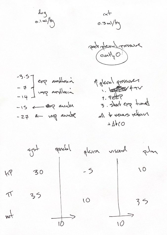
Pleural fluid forms a thin lubricating film. My note and textbook quote 0.1 mL/kg in dogs and 0.3 mL/kg in cats. These are textbook estimates, not validated reference intervals.
Under anaesthesia, the original pressure cues are −3.5 at expiration and −7 to −14 at inspiration. Awake, they are −15 at expiration and −27 at inspiration. All are cm H2O. The textbook's anaesthetised dog ranges are −3.4 to −6.8 at end expiration and −7 to −14.3 at peak inspiration. Its awake values come from telemetric study means. The note's “peak” heading does not make them universal peak values. Location, breathing, method and study conditions matter.
During positive-pressure ventilation, larger tidal volumes, PEEP or too little time to exhale can raise intrathoracic pressure. Incomplete expiration can leave trapped gas and intrinsic PEEP. Venous return and cardiac output can then fall. The effect depends on the animal and ventilation conditions.
Starling forces describe bulk fluid filtration. The lower drawing uses hydrostatic pressures of 30, −5 and 10, with colloid osmotic pressures of 35, 10 and 35 across systemic capillaries, pleural space and pulmonary capillaries. Its two net arrows are each 10 cm H2O, toward the right: into the space at the parietal side and out at the visceral side.
The textbook figure uses different values: pulmonary hydrostatic pressure 11 and colloid osmotic pressures 34, 8 and 34. Its net gradients are 9 and 10. These are model pressures, not fluid volumes or flow rates. We cannot subtract them to calculate fluid accumulation. Parietal lymphatics also help remove fluid.
References
Johnston & Tobias, 2nd ed., Ch. 22/105; Ednick et al. 2007; Pinsky 1984; Johnston et al. 1989.