
Control of alveolar ventilation
Read the draft explanation
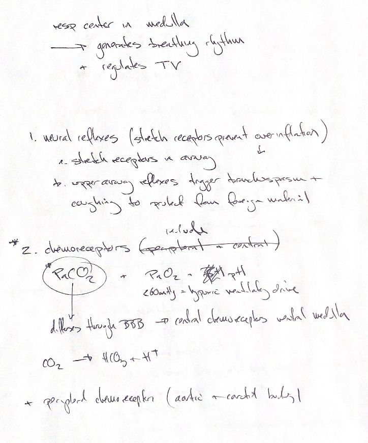
The respiratory network in the medulla sets the breathing rhythm and helps regulate tidal volume, the size of each breath.
Neural reflexes adjust that pattern. Stretch receptors in the airways and lung tissue can limit inspiration and help prevent overinflation. Irritants can trigger bronchospasm and coughing to protect the lungs from foreign material. Canine experiments show that sleep and arousal can alter these airway responses.
Chemoreceptors respond to PaCO2, PaO2 and pH. Pa means arterial partial pressure. CO2 is a major chemical signal. It readily crosses the blood-brain barrier and changes local H+ and pH in brain extracellular fluid. That helps stimulate central chemoreceptors, including those in the ventral medullary region. The chemical shorthand is CO2 + H2O ⇌ H+ + HCO3−. Bicarbonate movement into the brain also occurs; the barrier is not an absolute block to it.
Peripheral chemoreceptors in the carotid and aortic bodies also contribute. They respond to low oxygen and to changes in CO2 and pH. Central and peripheral signals work together.
The original note and textbook use PaO2 below about 60 mmHg as the cue for a stronger hypoxic ventilatory drive. The response is graded and depends on CO2 and the conditions being studied. It is not a sharp switch at one pressure or a treatment rule.
References
Johnston & Tobias, Veterinary Surgery: Small Animal, 2nd ed., Ch. 103; Johnson et al. 1983; Blain et al. 2009; Smith et al. 2015; Sullivan et al. 1979.