
Defecation reflex: pelvic, hypogastric and pudendal nerves
Read the draft explanation
Defecation combines enteric reflexes, autonomic nerves and conscious control. The internal anal sphincter is smooth muscle and works involuntarily. The external sphincter is striated muscle and allows voluntary control.
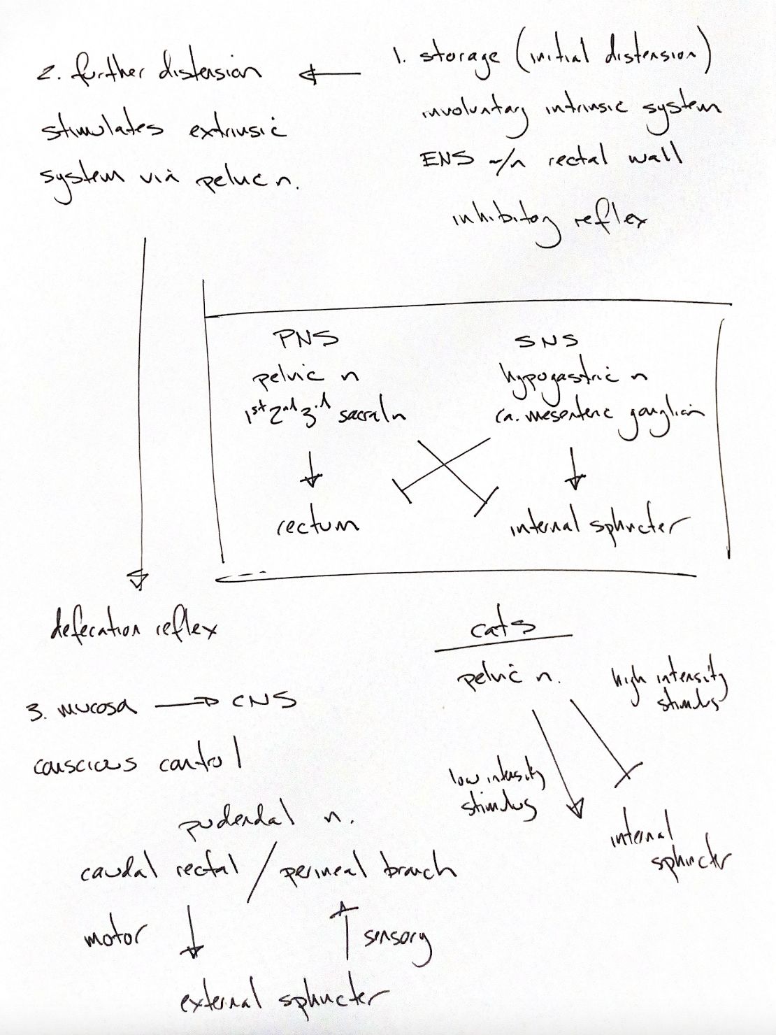
During storage, the rectum accommodates faeces. Distension also activates the enteric nervous plexus within its wall. The rectoanal inhibitory reflex relaxes the internal sphincter. With further distension, pelvic nerve pathways strengthen rectal contraction and internal-sphincter relaxation. These intrinsic and extrinsic mechanisms work together.
The pelvic nerves carry parasympathetic fibres from S1–S3. The basic pattern is excitation of the rectum and inhibition of the internal sphincter. Sympathetic fibres reach the pelvic plexus through the hypogastric nerves from the caudal mesenteric ganglion. Their usual pattern is the reverse: they inhibit rectal muscle and excite the internal sphincter. Experimental feline responses can be more complex.
Sensory input from the anal mucosa travels through pudendal pathways to the central nervous system. The caudal rectal branch of the pudendal nerve supplies motor input to the external anal sphincter. Perineal branches carry sensory input from the anal and perineal region. Conscious relaxation permits defecation; voluntary contraction can delay it.
The cat inset preserves an important experimental distinction. Low-intensity electrical pelvic nerve stimulation can contract the internal sphincter, while high-intensity stimulation can relax it. These findings came from anaesthetised cats. The response also depends on the existing anal tone and the neural pathways recruited. They do not define a clinical stimulation setting or a rule for every natural stimulus.
References
Johnston & Tobias, 2nd ed., Ch94; Carlstedt 1989; Buntzen 1996; Shafik 2002; University of Minnesota veterinary anatomy.