
Abdominal arterial supply
Read the draft explanation
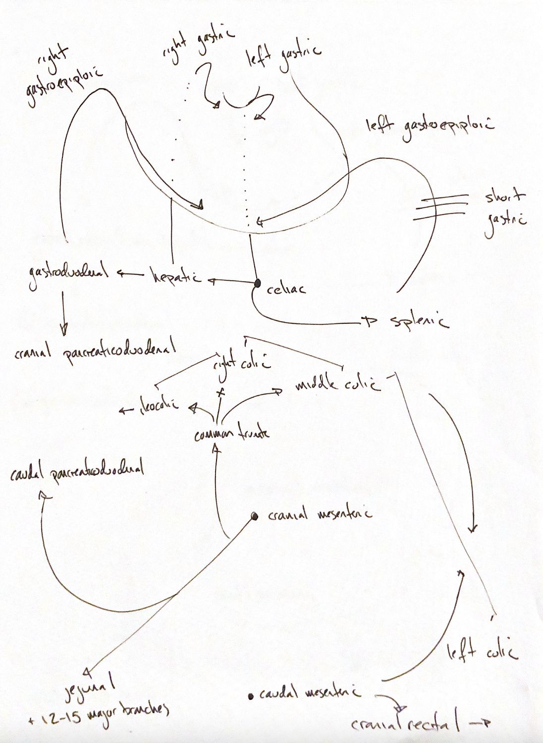
Start with three roots: the celiac, cranial mesenteric and caudal mesenteric arteries. This is a gastrointestinal branch map, not every abdominal vessel.
The celiac artery divides into hepatic, left gastric and splenic branches. The hepatic artery gives off the right gastric artery and continues as the gastroduodenal artery. This divides into the right gastroepiploic and cranial pancreaticoduodenal arteries.
The splenic artery supplies the short gastric arteries and continues as the left gastroepiploic artery. The right and left gastric arteries meet along the lesser curvature. The gastroepiploic pair meet along the greater curvature. Short gastric branches also connect with left gastric branches at the fundus. An anastomosis is a connection between vessels.
The cranial mesenteric artery gives off the caudal pancreaticoduodenal and jejunal arteries. My common trunk groups the ileocolic, right colic and middle colic arteries. This is a common canine pattern; origins vary, and cats often have separate colic branches.
My jejunal “12–15 major branches” is a canine textbook memory cue. The supplied book uses 12–15 for major cranial mesenteric branches, while other anatomy texts give different jejunal ranges. An exact primary canine sample was not verified. Branches form arcades, then short vasa recta reach the intestinal wall.
The caudal mesenteric artery gives a left colic branch running cranially and a cranial rectal branch running caudally. The left colic joins the middle colic. Cranial and caudal pancreaticoduodenal arteries also join, linking celiac and cranial mesenteric territories. Connections do not guarantee adequate collateral flow after a vessel is lost.
The curves, ticks and arrows are recall schematics, not fixed branch counts, vessel courses or measured flow.
References
Johnston & Tobias, Veterinary Surgery: Small Animal, 2nd ed.; UMN anatomy guide.