
Basic anatomy of the testes; testicular cells; accessory sex glands in dogs and cats
Read the draft explanation
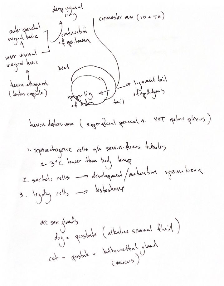
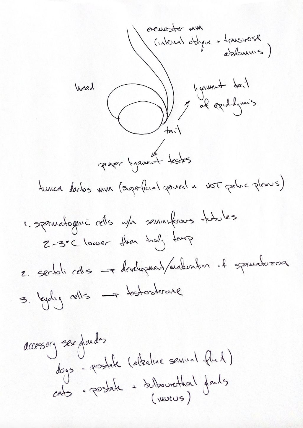
Start with the testis, its epididymis and their coverings. The epididymal head is cranial, the body follows the testis, and the tail is caudal. The proper ligament joins the tail to the testis. The ligament of the epididymal tail joins it to the vaginal tunic and spermatic fascia.
The outer parietal and inner visceral vaginal tunics continue from peritoneum. The vaginal cavity lies between them and communicates with the abdomen through the vaginal ring at the deep inguinal ring. The visceral layer coats the testis; the tunica albuginea forms its capsule underneath.
In the dog, the cremaster runs outside the parietal tunic. It comes mainly from internal abdominal oblique; transversus abdominis contributes occasionally. Dartos is smooth muscle in the scrotal wall. Its postganglionic sympathetic fibres travel with the superficial perineal nerve, rather than the pelvic plexus. Contraction draws the testes closer to the body.
Spermatogenic cells inside seminiferous tubules develop into spermatozoa. Sertoli cells support their development and maturation. Leydig cells sit between tubules and produce testosterone. The textbook temperature cue is about 2–3°C below body temperature for viable sperm production. Cremaster, dartos and pampiniform heat exchange help regulate that temperature.
The dog has the prostate as its accessory sex gland. The cat also has paired bulbourethral glands, which add thick mucus to seminal fluid. One source conflict matters here: the original and testis chapter call prostatic fluid “alkaline”, but the prostate chapter reports canine fluid pH 6.1–6.5, which is mildly acidic. Human prostatic-fluid pH should not be carried over to the dog.
References
Chris Wood’s two drawings; Johnston & Tobias, Veterinary Surgery: Small Animal, 2nd ed., chs 111 and 113; University of Minnesota dog/cat anatomy guide; Fair & Cordonnier 1978, PMID 32404 (human study; canine pH given as background).