
Bladder innervation
Read the draft explanation
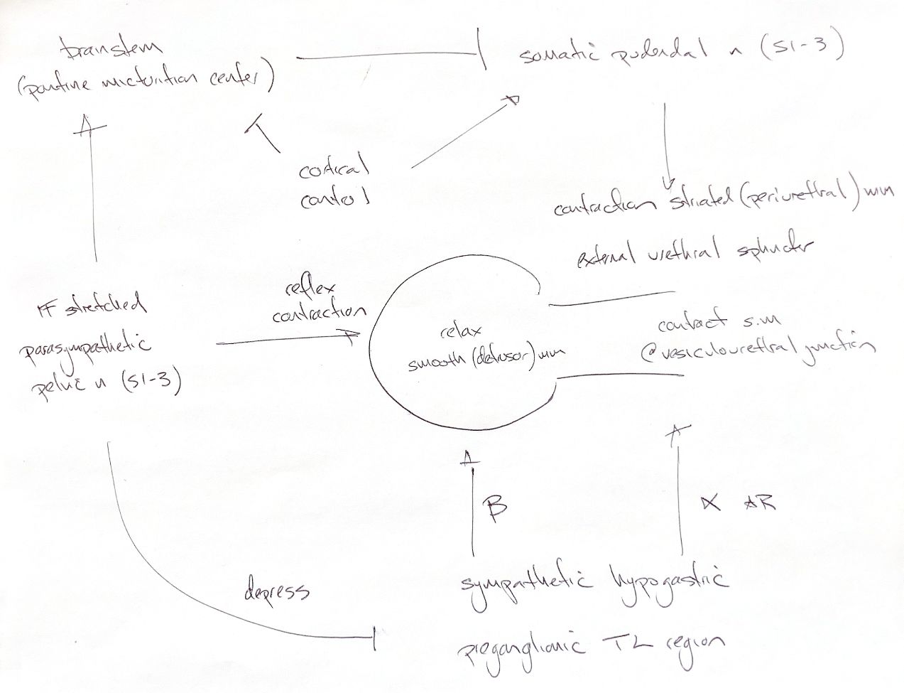
Storage and voiding use the same nerves in different ways. During storage, the sympathetic hypogastric nerves stimulate beta receptors in the detrusor. The bladder wall relaxes and holds more urine. At the same time, alpha-1 receptors in the bladder neck and proximal urethra raise smooth-muscle tone. The somatic pudendal nerve keeps the external striated urethral sphincter contracted. Its acetylcholine acts at nicotinic receptors.
As the bladder fills, stretch signals travel through the pelvic nerve to the sacral cord and ascend toward the brainstem. The parasympathetic pelvic pathway contracts the detrusor. The pontine micturition centre coordinates a sustained contraction with a relaxed outlet. Sympathetic outflow falls, so proximal urethral smooth-muscle tone falls. Pudendal motor activity also falls, allowing the external striated sphincter to relax. Pudendal stimulation itself contracts this muscle; it does not directly relax it. Cortical control helps decide when and where voiding occurs.
The original S1–S3 cue refers to spinal cord segments, not vertebrae. The supplied text uses S1–S3 for pelvic and pudendal pathways. The university anatomy guide places pudendal motor neurons mainly in S2–S3. Sympathetic levels also vary: the supplied prose gives L1–L4, its figure L1–L2, and the university guide L2–L4. The drawing therefore keeps the broader thoracolumbar label. The pontine centre coordinates the circuit; pelvic preganglionic neurons lie in the sacral cord.
“Internal sphincter” describes the functional smooth-muscle outlet. It is not a separate anatomical ring at the vesicourethral junction.
References
Chris Wood’s ACVS notes; Johnston & Tobias, Veterinary Surgery: Small Animal, 2nd ed., ch.32,116,118; University of Minnesota, Innervation of the Lower Urinary Tract.