
Nephron: functional unit and concentrating map
Read the draft explanation
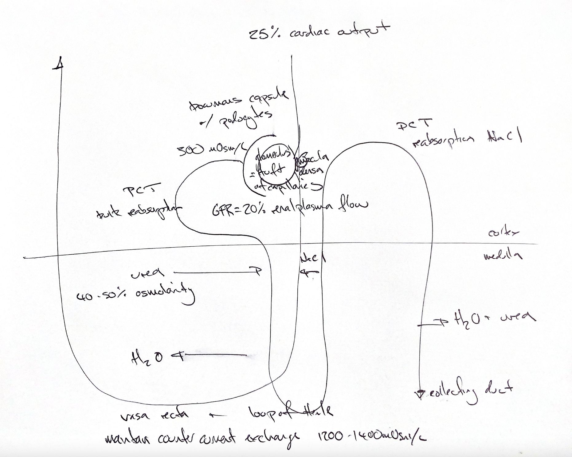
The nephron is the kidney’s functional unit. The overview is a route schematic. Follow the original map: the renal corpuscle sits in the cortex, the PCT loops to the left, Henle’s loop dips into the medulla, the DCT returns to the cortex and the collecting duct runs down on the right.
The glomerulus is a capillary tuft inside Bowman’s capsule. Podocytes cover the tuft as the capsule’s visceral layer. The macula densa belongs to the returning distal tubule beside the vascular pole. It senses tubular salt delivery and helps regulate filtration.
The original numbers are approximate textbook cues. The kidneys receive about 25% of cardiac output. Glomerular filtration rate is about 20% of renal plasma flow, which is different from whole-blood flow. Initial filtrate is about 300 mOsm/L.
The proximal convoluted tubule, or PCT, reabsorbs water and solutes in bulk. Water leaves the descending limb of Henle’s loop. NaCl leaves the ascending limb, especially its thick segment, with little water movement there. The distal convoluted tubule, or DCT, reabsorbs NaCl.
In the inner medullary collecting duct, ADH helps water leave the filtrate. Urea also leaves, enters the surrounding interstitium and recycles into a thin limb. The note’s 40–50% refers to urea’s contribution to medullary osmolarity. Its 1200–1400 mOsm/L medullary value is a textbook reference, alongside roughly 300 mOsm/L outside the medulla. Species and hydration alter this gradient; it is not a fixed canine urine ceiling.
The vasa recta are blood vessels alongside the loop. Descending blood loses water and gains solute; ascending blood gains water and returns solute to the medulla. This countercurrent exchange returns reabsorbed water to the circulation while limiting loss of the gradient. The tubular loop and urea handling help build the gradient; the vasa recta help preserve it.
References
Johnston and Tobias, Veterinary Surgery: Small Animal, 2nd ed., Chapter 114.