
Ligament tension and screw-home quick-reference
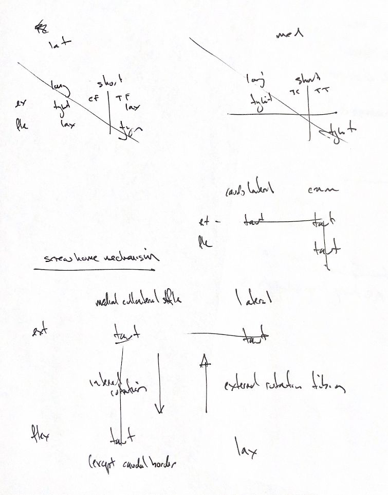
Read the draft explanation
Keep the joint name with each tension cue. The upper groups are the canine tarsal collaterals; the lower groups concern the stifle.
At the tarsus, both long collateral components are taut in extension and lax in flexion. Medially, the short tibiocentral part follows that pattern. The short tibiotalar part is lax in extension and taut in flexion. Laterally, the calcaneofibular part supports extension, while the talofibular part is lax in extension and taut in flexion.
Tension and total joint stability are different. The original sheet leaves calcaneofibular tension in flexion blank. A canine cadaver study found that the calcaneofibular and tibiotalar components contribute to static stability in both positions. The whole ligament complex, capsule and malleoli work together. A “lax” cue does not mean zero contribution to stability.
For the CCL, the craniomedial band stays taut in flexion and extension. The caudolateral band is taut in extension but lax in flexion. This is a gross functional model; individual fibres do not all carry identical tension.
At the stifle, both collateral ligaments are taut in extension. With flexion, most of the medial collateral remains taut, but its caudal border loosens. The lateral collateral becomes lax as its attachments move closer. This allows the lateral femoral condyle to move caudally relative to the tibia, coupled with internal tibial rotation.
Extension reverses that pattern. The lateral collateral tightens, the lateral femoral condyle moves cranially and the tibia rotates externally. This coupling is the screw-home mechanism. Rotation here means the tibia relative to the femur. In-vivo motion also depends on the activity, so this is a recall model rather than a fixed rotation angle.
References
Johnston and Tobias, Veterinary Surgery: Small Animal, 2nd ed., Chapters 61 and 64; Aron and Purinton 1985; Vasseur and Arnoczky 1981; Kim et al. 2015.