
Growth-plate zones
Read the draft explanation
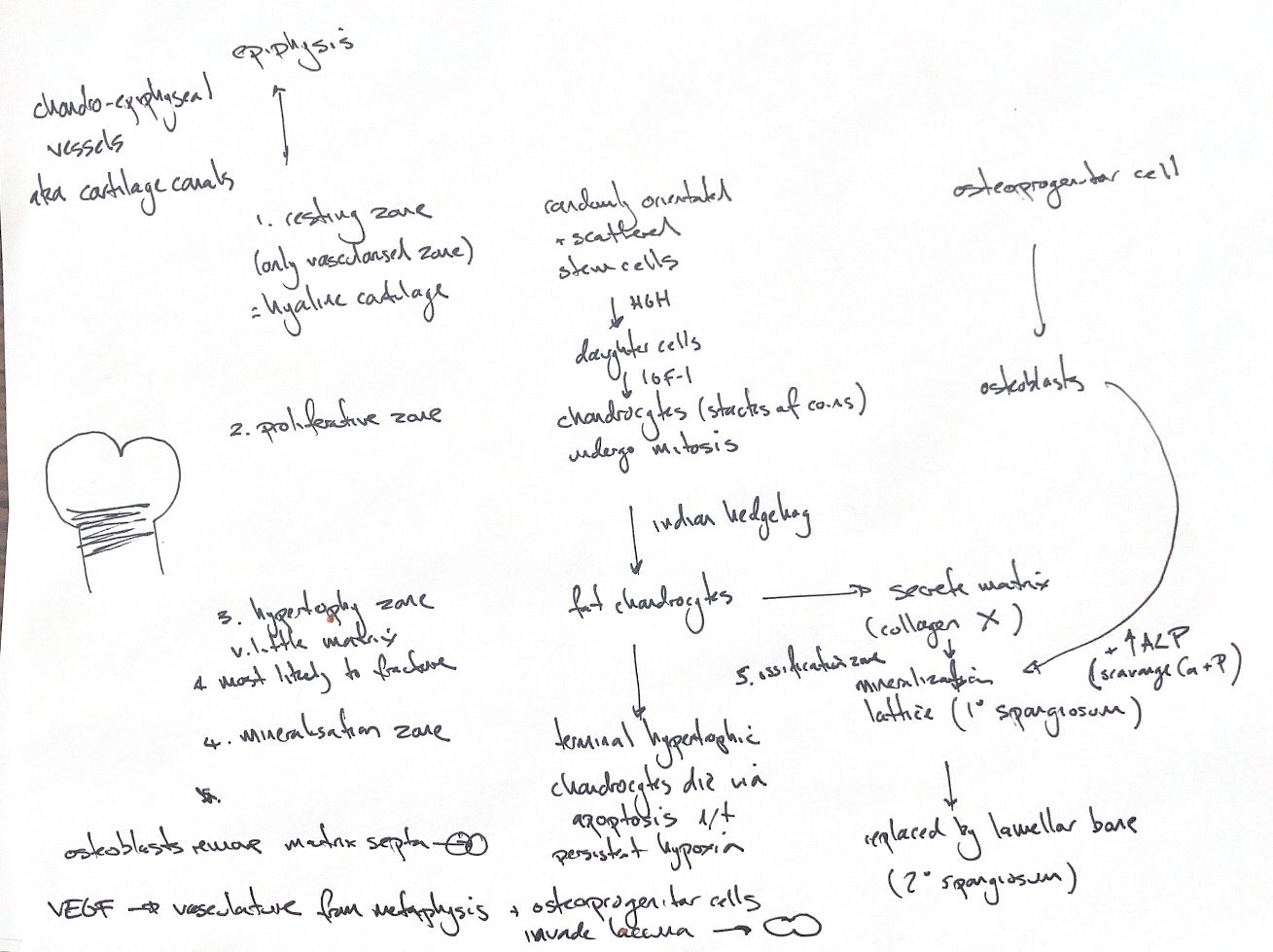
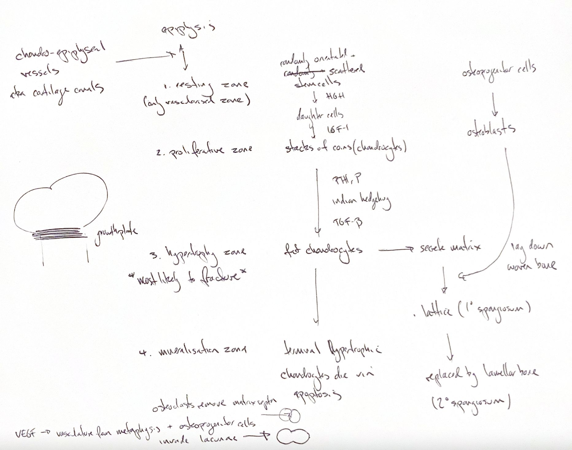
A growth plate turns cartilage into bone as a long bone lengthens. Its cartilage contains chondrocytes and type II collagen. Follow the cells from the epiphysis towards the metaphysis through five zones: resting, proliferation, hypertrophy, mineralisation and ossification.
Resting chondrocytes form a small, scattered stem-cell pool in hyaline cartilage. Chondro-epiphyseal vessels reach this region through cartilage canals. Growth hormone acts on the resting-cell population. Daughter cells produce IGF-1, which supports expansion of the proliferating chondrocytes.
In the proliferative zone, flat cells divide and line up like stacked coins. Local feedback involving PTHrP, Indian hedgehog and TGF-β regulates proliferation and progression towards hypertrophy. The hypertrophic cells enlarge and produce type X collagen. Little matrix remains between them, making this the weakest zone and a common plane for physeal fractures.
At the mineralisation front, terminal chondrocytes undergo apoptosis in the locally hypoxic matrix. Matrix vesicles contain alkaline phosphatase, or ALP, which supports calcium–phosphate mineralisation. Hypertrophic chondrocytes also produce VEGF, which promotes vascular ingrowth.
Clasts, including osteoclasts, remove transverse matrix septa. Vessels from the metaphysis and osteoprogenitor cells enter the empty lacunae. These cells become osteoblasts and lay woven bone on the remaining calcified cartilage scaffold. That lattice is the primary spongiosa. Remodelling replaces it with lamellar bone, the secondary spongiosa.
The five-zone scheme includes this ossification front. Vascular invasion here is distinct from the cartilage canals in the resting zone; the phrase “only vascularised zone” applies to the cartilage zones, not to newly forming metaphyseal bone.
References
hand-drawn ACVS Phase One study notes; Johnston and Tobias, Veterinary Surgery: Small Animal, 2nd edition, Chapters 39 and 73.