
Load-deformation and stress-strain
Read the draft explanation
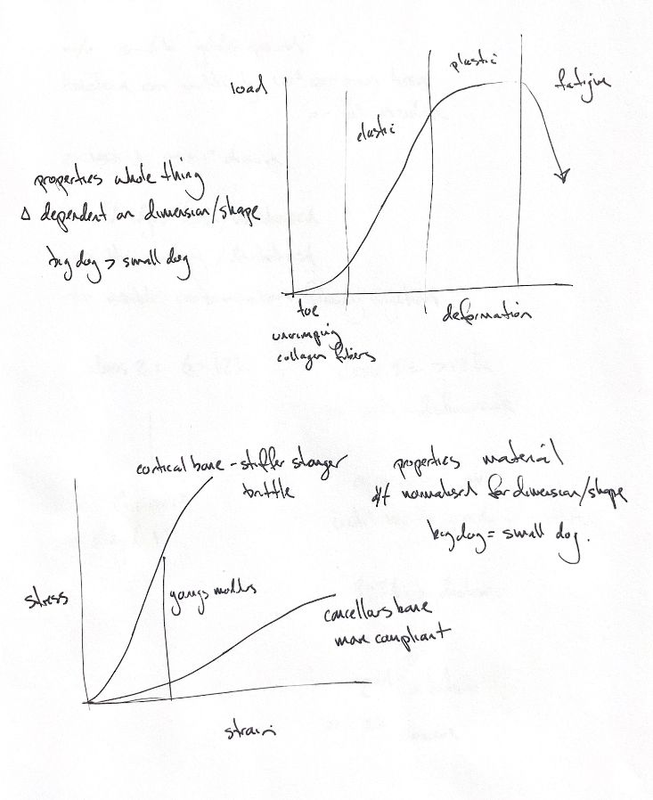
Load–deformation describes the whole specimen. Plot load on the vertical axis and deformation on the horizontal axis. Its slope gives structural stiffness. The result depends on material, size and shape, so a large dog’s bone and a small dog’s bone can behave differently as structures.
Stress–strain describes material behaviour. In a simple uniaxial test with a sufficiently uniform field, engineering stress is force divided by original area, and engineering strain is change in length divided by original length. This removes the direct area and length terms. It helps compare specimens, but it does not make different dogs’ tissues identical. Real bone has uneven stress and strain fields.
In collagen-rich tissue, the toe region reflects straightening of crimped fibres. The curve then becomes steeper as more fibres bear load. The elastic region permits recovery after unloading. Beyond yield, permanent deformation and damage develop. The specimen reaches its maximum load, then loses load-bearing capacity and fails. The curve is a teaching schematic; its exact shape depends on tissue and test conditions.
On a stress–strain plot, slope describes resistance to strain. Young’s modulus belongs to the ideal linear elastic model, with small deformation, homogeneity and isotropy assumed. For bone, the measured slope is better described as an apparent modulus under stated conditions. A load–deformation slope is structural stiffness, not Young’s modulus.
Under compression, cortical bone generally has greater apparent stiffness and strength than cancellous bone. It is also more brittle and tolerates less strain before failure. Cancellous bone is more compliant and can sustain greater strain. Its stiffness and strength depend strongly on density. Compare like loading directions, rates and specimen conditions.
References
Johnston & Tobias, Veterinary Surgery: Small Animal, 2nd ed., Ch39; Figs 39.18,39.20 and 39.25.