
Malignancy,metastasis and angiogenesis
Read the draft explanation
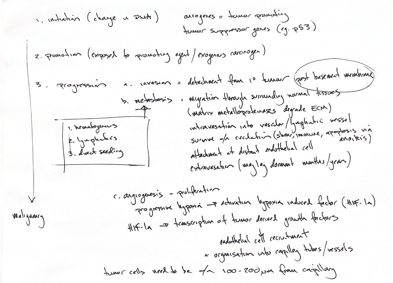
Initiation changes DNA. Oncogenes can drive growth signals, while loss of tumour suppressor function removes growth restraints. p53 is a key example. Promotion expands initiated cells; in chemical carcinogenesis, initiation comes before the promoting stimulus. Progression brings invasion, metastasis and angiogenesis.
To invade, cells detach from the primary tumour and cross the basement membrane. They migrate through surrounding tissue. Matrix metalloproteinases help break down the extracellular matrix. Intravasation puts cells into a blood or lymphatic vessel. They then face shear forces, immune attack and anoikis: apoptosis after losing matrix attachment.
A surviving cell can attach to distant endothelium, extravasate and enter new tissue. It may lie dormant for months or years. Later growth needs a suitable microenvironment, supporting stroma and blood supply. Tumours can spread through blood, lymphatics or direct seeding.
As growth outstrips oxygen supply, hypoxia activates HIF-1α. It promotes transcription of angiogenic signals such as VEGF. These recruit endothelial cells, which organise into capillary tubes and vessels. Blood flow supports growth and removes waste. The roughly 100–200 µm oxygen-diffusion scale varies with perfusion and oxygen use.
References
Johnston & Tobias, Veterinary Surgery: Small Animal, 2nd ed., ch. 25; Semenza 2008, doi: 10.1002/iub.93; Fidler 2001, doi: 10.1093/oxfordjournals.jncimonographs.a024251.