
Needle shape and suture attachment
Read the draft explanation
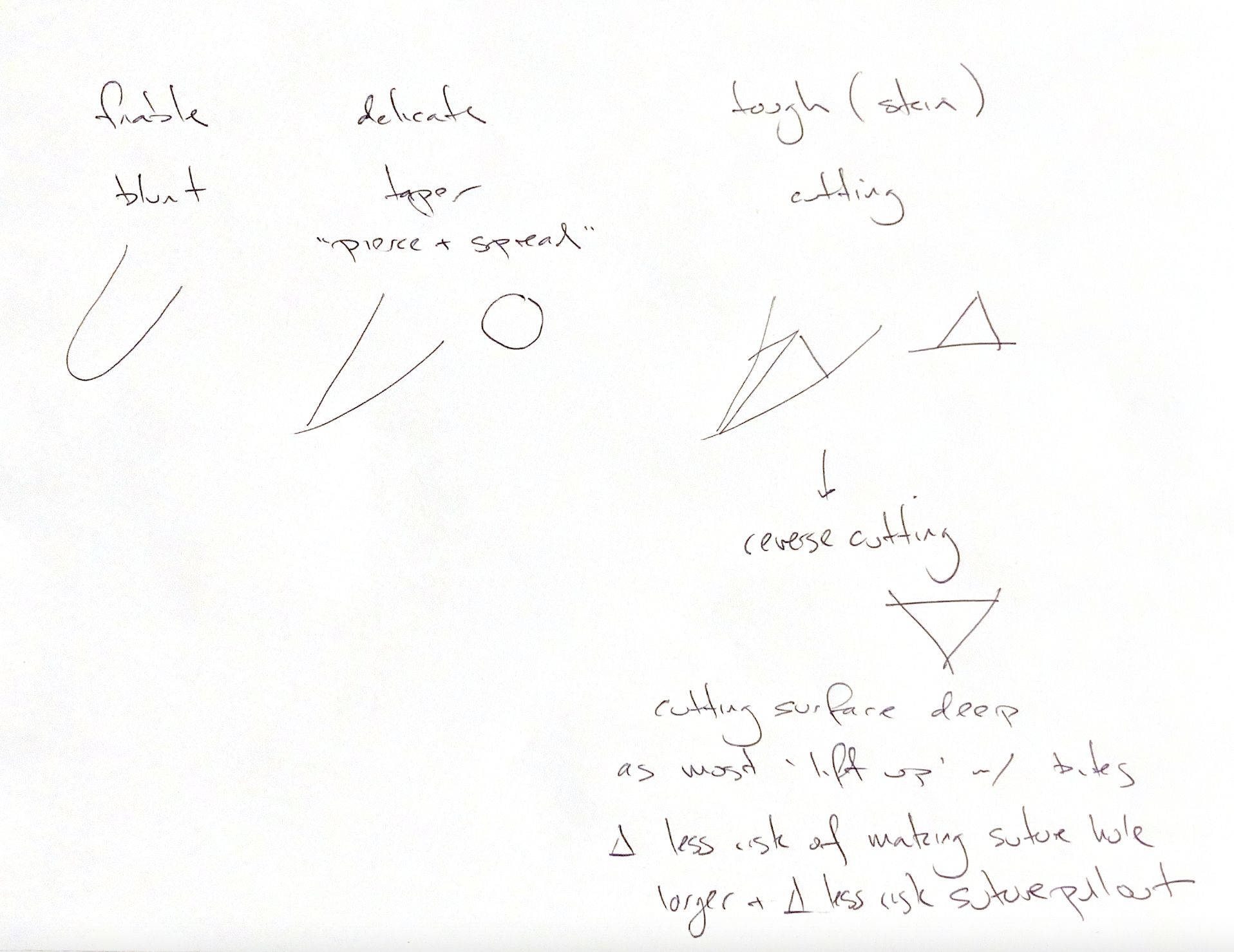
Match the point to the tissue. A blunt needle can part friable tissue, but it handles poorly in normal tissue. A taper point pierces and spreads delicate tissue. Its round point section helps keep the hole small in GI organs, fat, bladder and muscle.
Cutting needles pass more easily through tough tissue such as skin, fascia and periosteum. A conventional cutting needle has its cutting edge on the concave surface. It creates a triangular hole with the vertex toward the incision. Force toward the concavity can enlarge that hole and increase the risk of suture pull-through.
A reverse-cutting needle moves the cutting edge to the convex surface. The flat side of the triangular hole lies parallel to the incision. In theory, this lowers the risk of hole elongation and suture pull-through. Enter perpendicular to the tissue, then follow the needle’s curve. Pushing or pulling a curved needle straight enlarges the hole and adds trauma.
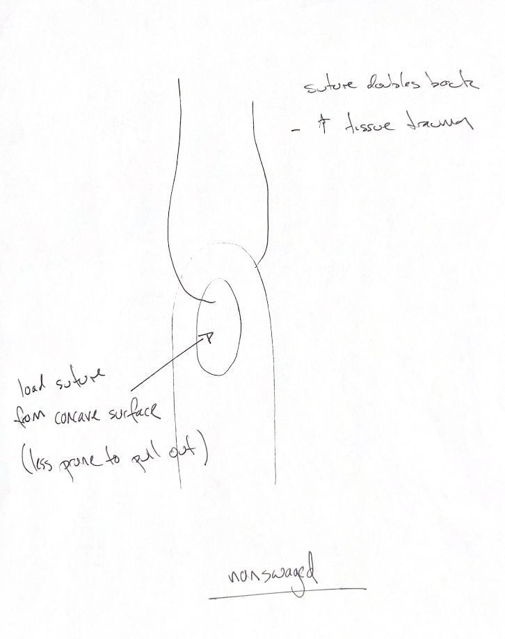
The attachment matters too. A swaged needle has no eye. The suture is fixed to the needle, creating a smaller attachment profile than an eyed needle. In a nonswaged needle, the suture passes through an eye and doubles back. This wider attachment opens a larger tract and causes more tissue trauma. Repeated use, cleaning and sterilisation can also dull these reusable needles.
Load an eyed needle from its concave surface. This makes the suture less likely to pull out of the eye. Unthreading at the eye and suture pulling through tissue are separate problems.
References
Schmiedt, “Suture Material, Tissue Staplers, Ligation Devices, and Closure Methods,” in Johnston & Tobias, Veterinary Surgery: Small Animal, 2nd ed., PDF pp. 597–599, Figs. 16.2–16.3.